Download
1 / 46
460 likes | 719 Vues
Chapter 13 The Respiratory System. Biology 112 Tri-County Technical College Pendleton, SC. System Essentials. Supply cells with oxygen Pick up carbon dioxide from the body Eliminate carbon dioxide from the body

E N D
Chapter 13 The Respiratory System Biology 112 Tri-County Technical College Pendleton, SC
System Essentials • Supply cells with oxygen • Pick up carbon dioxide from the body • Eliminate carbon dioxide from the body • Organs of respiratory system include: Nose, pharynx, larynx, trachea, bronchi and its branches, and the alveoli of the lungs
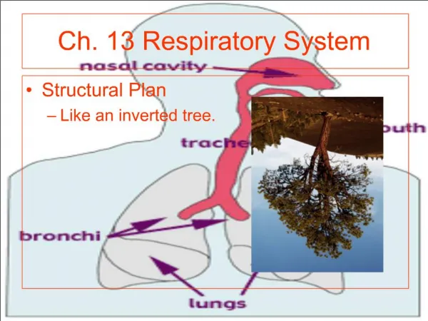
A nose for this…or that • Only externally visible part of system • Air enters nose through external nares (nostrils) • Interior consists of nasal cavity divided by midline nasal septum • Olfactory receptors in mucosa in superior part of cavity just below ethmoid bone
Nose, cont. • Rest of mucosa lining called respiratory mucosa rests on network of thin-walled veins that warms air flowing past • Mucosa moistens inhaled air & traps incoming debris • Ciliated cells move contaminated mucus posteriorly toward throat (pharynx) swallowed & digested by stomach acid
Nose, cont. • Three mucosa-covered lobes called nasal conchae increase surface area exposed to air • Also increase turbulence in nasal cavity • Nasal cavity separated from oral cavity by partition called the palate • Anterior palate supported by hard bone = hard palate • Posterior palate is unsupported by bone = soft palate
Paranasal Sinuses • Nasal cavity surrounded by paranasal sinuses located in frontal, sphenoid, ethmoid, and maxillary bones • Lighten the skull • Resonance chambers for speech • Produce mucus that drains into nasal cavity • Nasal mucosa continuous throughout RT • Nasal infections can spread throughout mucosa • Sinusitis difficult to treat & can cause marked change in voice quality
Pharynx • Muscular passageway for food and air commonly called throat • Naso-; Oro-; and laryngopharynx • Air enters superior portion (naso) from nasal cavity anteriorly • Air descends through oropharynx and laryngopharynx to enter larynx below • Food from mouth travels with air through oro- and laryngopharynx but directed posteriorly to esophagus instead of entering larynx
Pharynx, cont. • Auditory tubes that drain middle ear open into nasopharynx • otitis media (ear infection) may follow sore throat/pharyngeal infections • Clusters of lymphatic tissue called tonsils found in pharynx • Pharyngeal: (adenoids) located high in naso • Palatine: oropharynx at end of soft palate • Lingual: at base of tongue
Larynx and Associated Structures • Larynx (voicebox) routes air/food into proper channels and plays role in speech • Formed by 8 rigid hyaline cartilages and elastic cartilage called epiglottis • Largest of hyaline cartilages is THYROID cartilage (Adam’s apple) • Epiglottis protects superior opening of larynx
Larynx, cont. • Not swallowing, epiglottis does not restrict passage of air into respiratory passages • Swallowing, larynx pulls forward and epiglottis tips forming lid over opening of larynx • Routes food/drink into esophagus posteriorly • Cough reflexunconscious person
Larynx, cont. • Part of mucous membrane of larynx forms pair of folds called vocal folds (true vocal cords) which vibrate with expelled air • Allows speech • Slitlike passages between folds called glottis • Larynx leads to trachea (windpipe)
Trachea • Extends from larynx to level of 5th thoracic vertebra (~ midchest) • Lined with ciliated mucosa • Walls reinforced with C-shaped rings of hyaline cartilage • Open parts of rings abut esophagus and allow it to expand anteriorly when one swallows
Trachea, cont. • Solid portions support walls and keep it patent (or open) in spite of pressure changes during breathing • Tracheal obstructions are LIFE-threatening • Heimlich maneuver can unclog trachea • It works or my side would be gone…for good • Sometimes emergency trachesostomy is required
Bronchi Divisions • Trachea divides into right and left primary bronchi • Each primary bronchi plunges into medial depression (the hilus-depressed area where vessels enter/leave an organ) of lung • Right pulmonary bronchus is wider, shorter, and straighter than left • Smaller subdivisons of primary bronchi within lung deliver air to alveoli
Lungs and more… • Occupy most of thoracic cavity except for mediastinum (houses heart, great blood vessels, bronchi, esophagus, thymus, and trachea) • Narrow superior portion is APEX; located just deep to clavicle • Broad area resting on diaphragm is BASE • Each lung divided into LOBES by fissures • Left lung = 2 lobes; right lung = 3 lobes
Lungs, cont. • Surface covered by visceral (pulmonary) pleura and walls of thoracic cavity lined by parietal pleura • Membranes produce pleural fluid which allows lungs to glide easily over thorax wall during breathing • Also causes 2 pleural layers to cling together • Glide easily but resist being pulling apart • Absolutely essential for normal breathing
Lungs, cont. • PLEURISY (inflammation of pleura) can be caused by decreased secretion of pleural fluid • Surfaces become dry and roughfriction and stabbing pain with each breath • Another kind of pleurisy results in excess fluid and pressure on the lungs • Primary bronchisecondary & tertiary bronchibronchiolesterminal bronchioles (conducing zone structures)respiratory zone structures
Lungs, cont. • RZ structures include respiratory bronchiolesalveolar ductsalveolar sacsalveoli and is ONLY site of gas exchange
Exchange of Gases • Alveoli composed largely of single layer of layer of squamous epithelial cells • External surface of alveoli covered with cobweb of pulmonary capillaries • Alveolar and capillary walls construct respiratory membrane (air-blood barrier) • Blood flowing on one side; air on the other • Gases diffuse across air-blood barrier by simple diffusion
Gas Exchange, cont. • Surface area of lungs = size of racquetball court (70-80 sq. meters) • Macrophages (dust cells) wander in and out of alveoli to pick up bacteria/debris • Cuboidal epithelial cells scattered throughout alveolar walls secrete surfactant (lipid that coats gas-exchange alveolar surfaces and is important in lung function
Events of Respiration • Pulmonary ventilation (breathing) • External respiration (gas exchange between pulmonary blood and alveoli) • Respiratory gas transport (gas must be transported to/from lungs and tissue cells via bloodstream • Internal respiration (gas exchange between blood and tissue cells at systemic capillaries
Mechanics of Breathing • Pulmonary ventilation depends on volume changes in thoracic cavity • Volume changes lead to pressure changes flow of gases to equalize the pressure • Gas always fills its container • Volume of container related to pressure of gas • Inspiration = gas flowing into lungs • Expiration = gas flowing out of lungs
Mechanics of Inspiration • Diaphragm & external intercostal muscles contract • Size of thorax increaseslungs adhere tightly to thorax wallsstretched to new, larger size of thorax • Lung volume increases producing partial vacuum (pressure less than atmospheric pressure) • Air rushes in to fill space—inspiration is always an active process
Mechanics of Expiration • Usually passive process that essentially reverse of inspiration • Active expiration (forced)—internal intercostals activated and contracted to depress rib cage and abdominal muscles contract to help FORCE air from lungs • Asthma (spasms of bronchioles) or chronic bronchitis/pneumonia can narrow respiratory passageways
Mechanics, cont. • Actelectasis (lung collapse) renders lung useless for ventilation • air enters pleural space through chest wound or rupture of visceral pleural (allows air to enter pleural space from respiratory tract) • Pneumothorax is term given presence of air in intrapleural space (disrupts fluid bond between pleura)
Mechanics, cont. • RESPIRATORY SOUNDS: • Bronchial sounds produced by air rushing through large respiratory passageways such as trachea and bronchi • Vesicular breathing sounds occur as air fills alveoli • Soft and resemble muffled breeze
Modified Respiratory Movements • Situations other than breathing move air in and out of respiratory system • Most “nonrespiratory air movements” are result of reflex activity • Cough, sneeze, crying, laughing, hiccups, yawn, sighing….
External Respiration • Actual exchange of gases between alveoli and blood (pulmonary gas exchange) • Oxygen leaves alveolus and enters blood capillary • Carbon dioxide leaves blood capillary and enters alveolus • Occurs by simple diffusion (movement occurs toward area of lower [ ] of diffusing substance)
Gas Transport • Very small amount of O2 dissolved in blood • Most transported as oxyhemoglobin • Hb + O2 HbO2 • Most CO2 transported as bicarbonate ion • 20-30% carried inside RBCs bound to hemoglobin (at different site than oxygen) • Very small amount transported in plasma • CO2 + H2OH2CO3H+ + HCO3- • For carbon dioxide to diffuse out of blood into alveoli, reaction must be reversed
Internal Respiration • Gas exchange process that occurs between systemic capillaries and tissue cells • Carbon dioxide leaves tissues and enters blood • Oxygen leaves blood and enters tissues • All gas exchanges made according to the laws of diffusion
Respiration Controls • Activity of respiratory muscles, diaphragm, and external intercostals regulated by brain impulses carried by phrenic and intercostal nerves • Respiratory rhythm and depth control center located in medulla and pons • Medulla contains self-exciting inspiratory and expiratory centers • Sets the rhythm of breathing
Controls, cont. • Pons contains apneustic and pneumotaxic centers • Smooth out basic rhythms of inspiration and expiration • Work to maintain ~ 12-15 respirations/min. • EUPNEA= normal breathing rate • Inspiratory center active = inspire • Expiratory center active = expire • Apneustic center in pons=keeps inspiratory center going • Pneumontaxic center in pons=limits length of inspiration and promotes expiration
Rate/Depth Breathing Factors • PHYSICAL such as walking, coughing, and exercise • >body temp can > rate of breathing • CONSCIOUS such as singing, swallowing, or holding breath while swimming • voluntary control limited then involuntary takes over • EMOTIONAL such as fright, surprise, or “other”
Factors, cont. • CHEMICAL most important rate and depth factors • CO2 and O2 levels in blood • Increased levels of CO2 and decreased blood pH MOST important stimuli leading to increase in rate/depth of breathing • Changes in CO2 [ ] in blood act directly on medulla centers • Changes in O2 [ ] in blood detected by chemoreceptors in AORTIC ARCH and CAROTID body in carotid artery
Factors, cont. • Chemoreceptors send impulses to medulla when blood O2 levels are dropping • <s in O2 levels ONLY important when they are dangerously LOW • Increases rate/depth of breathing • Hypoventilation=accumulation of CO2 in blood and >ed blood acidity • Hyperventilation=CO2 removed from blood and <ed blood acidity • Acidosis or alkalosis can result
Respiratory Related Terms • Apena is cessation of breathing caused by hyperventilation • Dyspnea is labored or difficult breathing • Emphysema=alveoli enlarge as walls of adjacent chambers break through; chronic inflammation promotes fibrosis of lungs • Requires lots of energy to exhale • Chronic bronchitis=mucosa of lower respiratory passages become severely inflamed and produces extra mucus
Terms, cont. • Chronic bronchitis impairs ventilation and gas exchange and increases risk of lung infections • Pneumonitis=inflammation of alveoli of the lungs resulting in them becoming clogged with mucus and/or fluids
Volumes and Capacities • Tidal volume (TV) is amount of air moved in and out of lungs with each breath (500 ml) • Inspiratory Reserve Volume (IRV) is amount of air that can be forcibly taken in over tidal volume (2100-3200 ml) • Expiratory Reserve Volume (ERV) is amount of air that can be forcibly exhaled after tidal expiration (1200 ml)
Volumes, cont. • Residual Volume (RV) is amount of air left in lungs that cannot be voluntarily expelled • Vital Capacity (VC) is sum of TV + IRV + ERV • Total amount of exchangeable air • about 4800 ml in healthy young males • Dead space volume is air that remains in conducting zone