Download
1 / 62
630 likes | 639 Vues
1.1 – The structure and functions of the musculo-skeletal system. Learning objectives. To be able to describe the functions of the skeleton. To understand different bone classifications and functioning. To be able to recognise and label a skeleton.

E N D
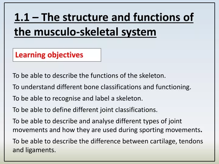
1.1 – The structure and functions of the musculo-skeletal system Learning objectives To be able to describe the functions of the skeleton. To understand different bone classifications and functioning. To be able to recognise and label a skeleton. To be able to define different joint classifications. To be able to describe and analyse different types of joint movements and how they are used during sporting movements. To be able to describe the difference between cartilage, tendons and ligaments.
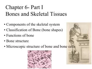
Functions of the skeleton The skeleton performs many functions in the body: Support – The skeleton supports the muscles. Protection – The skeleton protects delicate parts of the body like the brain. Muscle Attachment/Movement – Muscles are attached to the bones and move them creating levers. Blood cell production – blood cells are made in the bone marrow.
Support The skeleton acts as a framework. It gives the body support, enabling us to stand. The bones of the body are held together by ligaments. The skeleton provides a framework for the muscles, which are attached to bones by tendons.
Protection Some of our body parts, such as the brain, are very delicate and need protection. Bones can protect body parts from impactand injuries.
Muscle Attachment/Movement Muscles are firmly attached to bones forming levers which create sporting movements.
Blood cell production Long bones and other bones including the ribs, humerus, femur and vertebraebones, contain red bone marrow. This is where red blood cells are producedwhich carry oxygen. Other functions include:
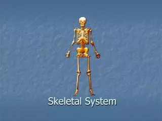
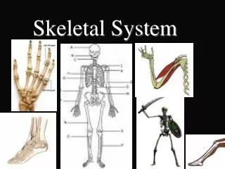
Skeletal System Without your skeleton you would be a shapeless sack of flesh. The adult skeleton has 206 bones. Watch me How many bones can you name?
Skeletal System Structure Cranium Clavicle Sternum Ribs Humerus Pelvis Radius Ulna Patella (knee cap) Femur Tibia Fibula
Skeletal System Structure Scapula Vertebral column Hand Carpals Metacarpals Phalanges Foot Tarsals Metatarsals Phalanges
The vertebral column It is made up of irregularly shaped bones called vertebrae. Between each vertebra there is a pad of cartilage which allows movement and prevents friction. The vertebrae protects the spinal cord. The vertebral column is divided into 5 sections.
Classification of bones Bones are divided into a number of different categories which have different roles in the body. 1. Long bones Long bones have a long shaft and are responsible for different types of movement. Sporting actions are created by long bones through levers. Long bones can be any size; they include the femur, humerus, tibia, fibula, metatarsals, metacarpals and phalanges.
2. Flat bones Flat bones perform a number of functions.
3. Short bones Carpals Short bones are light, small and very strong. The primary function is to support the weight of the body. The carpals in the wrist and the tarsals in the foot are examples of short bones. How does this aid sportspeople in an event? Gymnasts use carpals to support a handstand.
4. Irregular bones Femur Irregular bones are specially shaped to perform a particular function. Patella • These functions include: • Protection • Muscle attachment Tibia Fibula Examples include the patella and the vertebrae.
Classification of Joints DEFINITION: “A joint is a place where two or more bones meet”. Joints are responsible for the huge range of movement that the body can produce. There are several different types of joint classification.
Classification of Joints Hip 1. Ball and socket joint - the rounded end of a bone fits inside a cup-shaped end. Ball and socket joints allow movement in all directions. These are the most mobile joints in the body. Examples found in the body: Shoulders and hips. Why are these joints important for sport? Most sporting movements require movement by the shoulder and hip joints e.g. tennis serve
Classification of Joints 2. Hinge joints - only allow forwards and backwards movement like the hinge on a door. Examples found in the body: The knee and elbow. Why are these joints important for sport? These joints are extremely powerful and in conjunction with surrounding muscles can produce power and speed e.g. Knee drive during a 100m sprint
Classification of Joints 3. The Pivot joint has a ring of bone that fits over a pivoting bone. Pivot joints allow rotation only. Neck Examples found in the body: The joint between the atlas and axis in the neck allows turning and nodding of the head. Why are these joints important for sport? This joint allows for small movements that assist a larger sporting action e.g. breathing during a swimming stroke
Classification of Joints 4. Condyloid joints have an oval-shaped bone which fits into a similar shaped bone. They allow small movements in all directions. Examples found in the body: Found between the carpals and metacarpals in the wrist. Why are these joints important for sport? These joints are extremely useful when a sport involves gripping a ball. e.g. handball throw
Apply it! What has stuck with you? What synovial joints are used in these sporting examples?
Apply it! What has stuck with you?
Practice it! • Exam questions • 1. List three major types of bones, found in the human skeleton. (3) • Irregular • ___________________ • ___________________ • ___________________ • 2. Except for the femur, provide the names of two other bones in the leg that are classified as long bones. (2) • a) _______________________ • b) _______________________
Practice it! Exam questions 3. The humerus is a long bone. Which of the following statements correctly identifies a function of the humerus and its associated advantage to the performer in the statement? (1) A a hockey player can reach further to hit the ball as the humerus is a long bone B a footballer can kick the ball harder due to the length of the humerus C the humerus acts as a lever so a hockey player can apply more force to the ball D the humerus protects the footballer from injury.
Practice it! • Exam Questions: • 4.Name the bones of the upper and lower arm? (3) • 5.Name the anatomical name for the following bones (5) • a) Skull • b) Knee cap • c) Collar Bone • d) Shoulder blade • e) Wrist
Practice it! Exam Questions: 6. The following are regions or bones of the vertebral column. Place these regions of the vertebral column in the order they appear after the bones Atlas and Axis. (4) Atlas Axis 1 ......................................... 2 ......................................... 3 ......................................... 4 ......................................... Thoracic Sacral Atlas Lumbar Cervical Axis
Practice it! • Marks Scheme: • Short, Long, Flat • Tibia, Fibula • C • Humerus, Radius, Ulna • a) Cranium b) Patella c) Clavicle d) Scapula e) Carpals • Cervical, Thoracic, Lumbar, Sacral
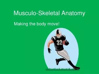
1.1 – The structure and functions of the musculo-skeletal system Learning objectives To understand the three muscle types and their functions To be able to label the voluntary muscles in our body To explain the term ‘antagonist pair’ and provide examples To understand the characteristics of fast and slow twitch muscle fibres
Connective tissues There are 3 types of connective tissue: Tendons connect muscles to bones. Ligaments are tough, elastic fibres that link bones to bones. Cartilage prevents the ends of bones rubbing together at joints.
Joint Movements 1. Flexion and Extension FLEXION – Decreasing the angle at a joint. (Bending the joint) EXTENSION - Increasing the angle at a joint. (Straightening the joint)
Joint Movements 2. Abduction and Adduction Abduction and Adduction is determined from the ‘MIDLINE’ of the body.
Joint Movements ADDUCTION – Sideways moving limb towards midline of the body. ABDUCTION – Sideways moving limb away from midline of the body REMEMBER: Adduction is to ADD towards the midline. REMEMBER: Abduction is to TAKE AWAY from the midline.
Joint Movements 3. Rotation/Circumduction The joint moves in a circular motion. e.g. Service action or bowling action.
Joint Movements 4. Planter-Flexion and Dorsi-Flexion Planter-flexion – The action of pointing toes away from the body. Dorsi-flexion – The action of pulling toes towards the body.
Apply it! What has stuck with you? What movements occur during these action?
Cyclist Footballer FLEXION – at the RIGHT knee joint EXTENSION - at the LEFT knee joint FLEXION – at the RIGHT hip joint as the leg raises FLEXION – Slight Flexion at the elbows FLEXION – Torso (body is bent forwards) EXTENSION – at the knee joints FLEXION – at the HIP JOINT of right leg ADDUCTION – at the hip joint as the left leg is moving towards the central line of the body ADDUCTION – Left arm FLEXION – at left elbow Swimmer - Start Butterfly Stroke ROTATION – at the shoulder joint EXTENSION – at elbow joints ABDUCTION – of the arms ADDUCTION – of the arms EXTENSION – at the knee joints EXTENSION – at the elbows
Label as many joint movements as you can see. i.e. flexion at the knee
Classification and Characteristics of Muscles Muscles are used in everyday life all the time. Sportspeople are reliant on the power of muscles to compete. What do you know about muscles already?
Classification and Characteristics of Muscles Muscles are involved in every movement in your body. Muscle is a special type of tissue made up of fibres that contract (shorten) and relax (lengthen). There are three types of muscle fibre.
Classification and Characteristics of Muscles 1. Voluntary Muscles These are attached to bones and they work whenever we want them to. e.g. Biceps & Triceps. These muscles are under our conscious control.
Classification and Characteristics of Muscles 2. Involuntary muscle These are found on the walls of the internal organs and they contract in waves. Food travels through the digestive system and blood through the blood vessels in this way. It works without you consciously controlling it, or even being aware of it.
Classification and Characteristics of Muscles 3. Cardiac muscle This is a special type of muscle that forms the walls of the heart chambers. It is a type of involuntary muscle, as it contracts without conscious thought or effort. It works non-stop without ever tiring. When it contracts it pumps blood out of the heart and around the body.
Classification and Characteristics of Muscles All three types of muscle are important in physical activity:
Voluntary Muscles Triceps Pectorals Deltoid Bicep LatissimusDorsi External Obliques Abdominals Gluteus Maximus Hip Flexors Hamstring Quadriceps Gastrocnemius Tibialis Anterior
Voluntary Muscles What happens when muscles contract? Muscles shorten when they contract and lengthen when they relax.
Voluntary Muscles When you contract your QUADRICEP what is the effect on the limb? When you contract your HAMSTRING what is the effect on the limb? When you contract your DELTOID what is the effect on the limb?
Now complete the grid below. What happens when each muscle contracts?
Raises arm sideways at the shoulder (abduction) Bends arm at the elbow (Flexion) Flexes trunk so you can bend forward Straighten leg at the knee (extension) Draws arm across chest Pulls arms backwards towards back Allows twisting and turning of the torso Straightens arm at the elbow joint (extension) Pull leg back at hip. Bends leg at the knee. (flexion) Flexes the ankle joint so you can pull toes towards the body Extends the ankle joint so you can stand on tiptoes Pulls upper leg towards the chest
Antagonistic muscle action The fixed or non-moving end is known as the origin. The insertion is known as the moving end. Muscles are arranged in antagonistic pairs. As one muscle contracts (shortens) its partner relaxes (lengthens). E.g. Bicep and Tricep.
Antagonistic muscle action Can you think of another antagonists pair in the body?
Antagonistic muscle action Gastrocnemius and Tibialis Anterior acting at the ankle joint Hamstring and Quadricep acting at the knee joint. Hip Flexors and Gluteus Maximus acting at the hip joint.