Download
1 / 62
1.13k likes | 2.37k Vues
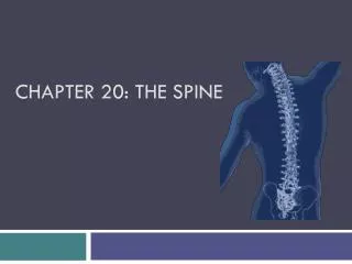
Chapter 25 The Thoracic Spine. Review of Anatomy Typical Thoracic Vertebrae. Atypical Anatomy. Five of twelve vertebrae are considered atypical (T1 and T9-T12). Most apparent difference between thoracic spine and remainder of the spine are the twelve ribs and their articulations.

E N D
Atypical Anatomy • Five of twelve vertebrae are considered atypical (T1 and T9-T12). • Most apparent difference between thoracic spine and remainder of the spine are the twelve ribs and their articulations.
Typical Thoracic Vertebrae (T2-T8)Body and Intervertebral Joint • Ratio of disk:vertebral body height – Less in thoracic spine than in cervical or lumbar regions. • Ratio of disk diameter:disk height – 2–3 times higher in thoracic spine than in lumbar spine. • Acute angular orientation of lamellae of anulus and small nucleus pulposus. Clinical Significance Creates stiffness and stability
Spinous/Transverse Processes • Slope inferiorly and overlap spinous processes of adjacent inferior vertebrae. Limits extension • Facet articulates with tubercle of rib to form costotransverse joint on ventral aspect. Restricts motion of rib in rotation about an axis parallel to and through neck of rib.
Transverse Processes • Upper and mid thoracic spine (T1-T6) facet is concave, corresponding to convex tubercle on neck of rib. • Facet is planar in lower thoracic region (T7-T10). Shape of lower thoracic costotransverse joints allows rib more flexibility during respiration and motion of thorax.
Facets • Orientation of the zygapophyseal joints (ZJ) depends on the region of the thorax. ZJ orientation guides and restricts mobility. • Posterolateral corners of superior and inferior aspects of vertebral body contain ovoid demifacet (except T1, T11, and T12). Development of costovertebral joint delayed until early adolescence, contributing to flexibility of the young thorax.
T1 Superior costal facets are circular to articulate with head of 1st rib. Spinous process is horizontal and is long and prominent as C7. T9 Inferior costal facets are absent and there is no direct articulation with the 10th ribs. T10 No inferior costal facets and no direct articulation with the 11th ribs. Atypical Vertebrae (T1, T9, T10)
T11 Articulates only with heads of 11th ribs. Transverse processes are small and do not have articular facets for tubercles of ribs. T12 Possesses only two costal facets for the 12th ribs. Body, transverse processes, and inferior facets are similar to lumbar vertebrae. Atypical Vertebrae (T11, T12)
Ribs • Ribs 1–7 – True ribs • Ribs 8–10 – False ribs • Ribs 11, 12 – Floating ribs Rib Functions: • Protect heart, lungs, and great vessels against trauma • Provide attachment for skeletal and respiratory muscles • Facilitate postural alignment and upper extremity function
Kinetics ROM • Flexion and extension – More limited in upper thoracic region (facets lie closer to frontal plane). Flexion – 20–45 degrees Extension – 20–45 degrees • Lateral flexion increases in lower thoracic region. Lateral flexion 20–40 degrees
Kinetics (cont.)ROM • Rotation is more limited in lower thoracic region. Rotation 35–50 degrees in each direction Lee states: If lateral flexion in frontal plane occurs first it is accompanied by contralateral rotation BUT if rotation in transverse plane occurs first it is accompanied by ipsilateral rotation. Lee DG. Manual Therapy for the Thorax – A Biomechanical Approach. Delta, British Columbia, Canada: DOPC, 1994.
Respiration During Inhalation • Pump handle movement is result of anterior aspect of rib moving superiorly. • Bucket handle movement is result of lateral aspect of rib moving superiorly. During Exhalation • Anterior and lateral aspects of ribs move inferiorly.
Extension Spinalis capitis, cervicis, thoracis Longissimus thoracis Semispinalis thoracis Rotatores thoracis Multifidus Interspinales Flexion Levatores costarum Rectus abdominis Internal obliques External obliques Myology of Thoracic Spine
Lateral Flexion Longissimus thoracis Iliocostali thoracis Semispinalis thoracis Multifidus Intertransversarii Levatores costarum Rotation Iliocostalis thoracis Semispinalis thoracis Rotatores thoracis Multifidus Intertransversarii Internal obliques External obliques Levatores costarum Myology of Thoracic Spine (cont.)
Rib Depression Longissimus thoracis Iliocostalis lumborum Rib Elevation Iliocostalis cervicis Viscera compression Transversus abdominis Respiration Diaphragm (inspiration) Intercostals (inspiration/expiration) Rectus abdominis (expiration) Internal/external obliques (expiration) Transversus abdominis (expiration) Myology of Thoracic Spine (cont.)
Inspiration Levatores costarum Pectoralis major/minor Rhomboids Anterior/medial/posterior scalenes Serratus anterior and posterior superior Subclavius, SCM Thoracic erector spinae Trapezius Expiration Iliocostalis lumborum Transversus thoracis Inspiration/Expiration Latissimus dorsi Quadratus lumborum Serratus posterior inferior Maintenance of rib cage shape Intercostals Accessory Muscles of Respiration
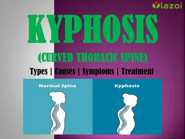
Anatomic ImpairmentsKyphosis An exaggeration of the normal posterior curve of the spine. • Results from change in structure and shape in spine or posture. • Fracture of anterior aspect of vertebral body – Osteoporosis (OP). • Scheuermann’s disease – Hereditary disorder that results in kyphosis.
Osteoporosis Low bone density, skeletal fragility, and fracture. Intervention • Consult with referring provider to determine if fracture is stable. • Pain control – Medications, back braces, and physical therapy modalities. • Moderate weight-bearing exercise (e.g., walking). • Resisted upper extremity exercise. • Balance training exercises.
Scheuermann’s Disease At least three wedged adjacent vertebral bodies of five degrees or more. Intervention • Usually limited to patients with painful deformity, painful progression, and at least two years of growth remaining. • Manage with bracing until skeletal maturity. • Strengthen spinal extensors. • Stretch hamstrings, pectoralis major, superior rectus abdominus, and anterior longitudinal ligament.
Scoliosis – 3 Types Lateral curvature of the spine, involving lateral flexion and rotation of the involved region(s). 3 Types: • Nonstructural scoliosis • Transient structural scoliosis • Structural scoliosis (idiopathic accounts for 70–80% of cases of scoliosis)
Examination and Evaluation • History • Systems review • Disorders of other systems can mimic thoracic pain (i.e., cancer, vascular disease, etc.). • Skeletal systems review – Scan examination of both upper and lower quadrants. • Elderly females with kyphosis screened for OP. • Individuals with exaggerated thoracic stiffness screened for ankylosing spondylitis. Refer to appropriate healthcare provider when indicated!
Aerobic capacity Ergonomics and body mechanics Gait, locomotion, balance Joint mobility, integrity Motor function Muscle performance Pain tests and disability measures Posture ROM and muscle length Sensory integrity Ventilation, respiration, and circulation Additional medical screening (radiographs, etc.) Choice of tests depends on results of history and systems review. Tests and Measures
Therapeutic Exercise for Common Physiologic Impairments Impaired Muscle Performance Sources: • Neurologic impairment or pathology • Muscle strain or injury • Disuse resulting in atrophy and general deconditioning • Length-associated changes resulting in altered length-tension properties
Neurologic Impairment or Pathology Treatment • Neural input must be restored for muscle performance to improve. • Protect weakened muscles from overstretch with proper support. • Stretch short muscles to maintain extensibility and prevent contracture. • For example: Impaired respiration – Stretch short muscles and apply manual or elastic band resistance to facilitate strength.
Muscle Strain or Injury • Address posture and movement patterns. • Improve performance of underused synergists. • For example, in the case of overuse of anterior scalene during breathing, reduce anterior scalene use by improving performance of deep anterior cervical flexors and instruct in proper pump and bucket handle diaphragmatic breathing.
Disuse Resulting in Atrophy and General Deconditioning Caused by illness, immobilization, sedentary lifestyle, subtle shifts in muscle balance. • Progressive resistive exercises for the upper body. • Initially, weight of limb is ample stimulus. • Progress in small increments. • Address balance between abdominal and spinal extensors as well as thoracic multifidii.
Length-Associated Changes Subtle imbalances in muscle length lead to length-associated strength changes and positional weakness of one synergist compared with agonist or antagonist. • Strengthen weak overstretched muscle groups in shortened range. • Stretch adaptively shortened muscles. • Supportive taping adjunctive. • Correction of posture and movement patterns.
Impaired ROM, Muscle Length, and Joint Mobility/Integrity • Optimal function of the thoracic region requires full symmetrical cardinal plane motion and full rib motion. • Consider symmetrical breathing patterns. • Diagnose restrictions that are joint versus soft tissue origin.
Hypermobility • First, determine contributing impairments. • Improve muscle balance and stability of trunk musculature (i.e., superficial vs. deep, anterior vs. posterior). • Consider effect of kinematic chain from ground upward (i.e., foot, ankle, knee, hip, pelvis). • Improve motor control (e.g., hold spine in ideal alignment during movements of extremities). • Improve mobility of adjacent hypomobile segments/regions. • Prevent thoracic flexion through use of bracing or taping.
Hypomobility • First, establish contributing impairments to hypomobility. • Establish need for joint and/or soft tissue mobilization. • Include passive stretching, AROM exercise. • Stabilize mobile segments while stretching hypomobile segments.
Muscle/Myofascial Length Treatment • Specific soft-tissue mobilization followed by exercises to maintain new mobility. • Passive stretch with diaphragmatic breathing for restrictions in oblique abdominal length. • As stability/mobility progresses – Progress to full arcs of motion.
Impaired Posture and Motor FunctionKyphosis • Manual and soft tissue mobilization • Self-mobilization • Manual stretching of pectoralis major/minor, intercostals, lumbar spine extensors, shoulder adductors • Tape thoracic spine for feedback • Strengthen thoracic extensors and cervical spine flexors
ScoliosisCorrection of asymmetrical postural habits (prevention during childhood)
Lordosis Treatment • Improve impairments of shoulder girdle. • Modify traditional exercises to prevent thoracic extension. • Self-mobilization techniques (promoting thoracic flexion and rotation).
Modified Middle and Lower Trapezius Strengthening for Individuals with Thoracic Lordosis