Download
1 / 52
520 likes | 727 Vues
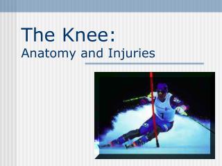
Chapter 20: The Knee and Related Structures. Jennifer Doherty-Restrepo, MS, LAT, ATC Academic Program Director, Entry-Level ATEP Florida International University Acute Care and Injury Prevention. Assessment of the Knee Joint. Determine MOI - This is critical!!! History: Acute Injury

E N D
Chapter 20: The Knee and Related Structures Jennifer Doherty-Restrepo, MS, LAT, ATC Academic Program Director, Entry-Level ATEP Florida International University Acute Care and Injury Prevention
Assessment of the Knee Joint • Determine MOI - This is critical!!! • History: Acute Injury • Past history • Position of body at time of injury? • Did the knee collapse? • Did you hear or feel anything? • Could you move your knee immediately after injury or was it locked? • Did swelling occur? • Where was the pain?
History: Recurrent or Chronic Injury • What is your major complaint? • When did you first notice the condition? • Is there recurrent swelling? • Does the knee lock or catch? • Is there severe pain? • Grinding or grating? • Does it ever feel like giving way? • What does it feel like when ascending and descending stairs? • What past treatment have you undergone?
Observation • Walking, half squatting, going up and down stairs • Swelling, ecchymosis • Leg alignment • Genu valgum and genu varum • Hyperextension and hyperflexion • Patella alta and baja • Patella rotated inward or outward • Tibial torsion, femoral anteversion and retroversion
Observation cont. • Knee Symmetry or Asymmetry • Do the knees look symmetrical? • Is there obvious swelling? • Atrophy? • Leg Length Discrepancy • Anatomical or functional • Anatomical differences can potentially cause problems in all weight bearing joints • Functional differences can be caused by pelvic rotations or mal-alignment of the spine
Palpation - Swelling • Intracapsular swelling • May be referred to as joint effusion • Swelling within the joint that is caused by synovial fluid and blood is called hemarthrosis • Sweep maneuver – sign of joint effusion • Ballotable patella - sign of joint effusion • Extracapsular swelling • Localized over the injured structure • May ultimately migrate down to foot and ankle
Medial Collateral Ligament Sprain • MOI = severe blow or outward twist • Grade I: Signs and Symptoms • Little fiber tearing or stretching • Stable valgus test • Little or no joint effusion • Some joint stiffness and point tenderness on lateral aspect of the knee • Relatively normal ROM
Grade I: Management • RICE for 24 hours • Crutches if necessary • Rehab • Cryokinetics • Isometrics • Progress to SLRs, bicycle riding, and isokinetics • Return to play when all areas have returned to normal • May require 3 weeks to recover
Grade II: Signs and Symptoms • Complete tear of deep capsular ligament and partial tear of MCL • No gross instability; laxity at 5-15 degrees of flexion • Slight swelling • Moderate to severe joint tightness • Decreased ROM • Pain along medial aspect of knee
Grade II: Management • RICE for 48-72 hours • Crutch use until acute inflammation phase has resolved • Possibly a brace or casting prior to the initiation of ROM activities • Modalities 2-3 times daily for pain • Gradual progression from isometrics (quad exercises) to CKC exercises; functional progression activities
Grade III: Signs and Symptoms • Complete tear of supporting ligaments • Complete loss of medial stability • Minimum to moderate swelling • Immediate pain followed by ache • Loss of motion due to effusion and hamstring guarding • Positive valgus stress test
Grade III: Management • RICE • Conservative non-operative versus surgical approach • Limited immobilization (with a brace) • Progressive weight bearing and increased ROM over 4-6 week period • Rehab would be similar to Grade I & II injuries
Lateral Collateral Ligament Sprain • MOI = Varus force usually with the tibia internally rotated • Direct blow is rare MOI • If severe enough damage may also occur to • Cruciate ligaments • ITB • Meniscus • Bony fragments may result as well
Signs and Symptoms • Pain and tenderness over LCL • Swelling and effusion around the LCL • Joint laxity with varus testing • May cause irritation of the peroneal nerve • Management • Same as MCL injury management
Anterior Cruciate Ligament Sprain • MOI = tibia externally rotated with a valgus force • Occasionally the result of hyperextension resulting from a direct blow • Research is quite extensive in regards to impact of femoral notch, ACL size and laxity, mal-alignments (Q-angle), and faulty biomechanics • Extrinsic factors may include, conditioning, skill acquisition, playing style, equipment, preparation time • May also involve damage to other structures including meniscus, capsule, and MCL
Signs and Symptoms • Experience pop with severe pain and disability • Positive anterior drawer and Lachman’s • Rapid swelling at the joint line • Other ACL tests may also be positive • Management • RICE; use of crutches • Arthroscopy may be necessary to determine extent of injury • Surgical repair • Without surgery, joint degeneration may result • Surgery may involve joint reconstruction with grafts (tendon), transplantation of external structures • Also requires 4-6 months of rehab
Posterior Cruciate Ligament Sprain • MOI = fall on bent knee (most common) • Most at risk during 90 degrees of flexion • Injury may result due to a rotational force • Signs and Symptoms • Feel a pop in the back of the knee • Tenderness and relatively little swelling in the popliteal fossa • Laxity with posterior sag test
Management • RICE • Non-operative rehab • Appropriate for grade I and II injuries • Focus on quad strengthening • Post-operative rehab • Surgery will require 6 weeks of immobilization in extension • Full weight bearing on crutches • ROM after 6 weeks • PRE at 4 months
Meniscal Lesions • Most common MOI is rotary force with knee flexed or extended • Tears may be longitudinal, oblique, or transverse • Medial meniscus is more commonly injured due to ligamentous attachments and decreased mobility • Also more prone to disruption through torsional and valgus forces
Signs and Symptoms • Effusion developing over 48-72 hours • Pain in joint line • Loss of motion • Intermittent locking and giving way • Pain with squatting • Portions of meniscus may become detached causing locking, giving way, or catching within the joint • If chronic injury, recurrent swelling or muscle atrophy may occur
Management • No locking but indications of a tear are present • Further diagnostic testing may be required • If locking occurs, anesthesia may be necessary to unlock the joint • Possible arthroscopic surgery • Healing dependent on location of tear • Menisectomy • Partial weight bearing, quick return to activity • Repaired meniscus • Requires immobilization, gradual return to activity over the course of 12 weeks
Knee Plica • MOI = irritation of the plica • Often associated with chondromalacia • Signs and Symptoms • Possible history of knee pain/injury • Recurrent episodes of painful pseudo-locking • Possible snapping and popping • Pain with stairs and squatting • Little or no swelling • No ligamentous laxity • Management • Treat conservatively w/ RICE and NSAID’s if the result of trauma • Recurrent conditions may require surgery
Osteochondritis Dissecans • MOI = partial or complete separation of articular cartilage and subchondral bone • Exact cause is unknown but may include: • Blunt trauma, • Possible skeletal or endocrine abnormalities, • Prominent tibial spine impinging on medial femoral condyle, or • Impingement due to patellar facet
Signs and Symptoms • Aching pain and point tenderness • Recurrent swelling • Possible locking • Possible quadriceps atrophy • Management • Rest and immobilization for children • Surgery may be necessary in teenagers and adults • Drilling to stimulate healing, pinning, or bone grafts
Loose Bodies • MOI = repeated trauma • May result due to osteochondritis dissecans, meniscal fragments, synovial tissue damage, or cruciate ligaments injury • Signs and Symptoms • May become lodged and cause locking or popping • Pain • Sensation of instability • Management • If not surgically removed it can lead to conditions causing joint degeneration
Joint Contusions • MOI = direct blow • Signs and Symptoms • Severe pain • Acute inflammation • Loss of movement • Swelling • If not resolved within a week then a chronic condition may exist (synovitis or bursitis) • Ecchymosis • Possible capsular damage • Management • RICE • Progress to normal activity following return of ROM • Padding for protection
Peroneal Nerve Contusion • MOI = compression due to a direct blow • Signs and Symptoms • Local pain and possible shooting nerve pain • Numbness and paresthesia • Added pressure may exacerbate condition • Generally resolves quickly • In the event it does not resolve, it could result in drop foot • Management • RICE • Return to play once symptoms resolve and no weakness is present • Padding for fibular head
Bursitis • MOI = acute, chronic, or recurrent swelling • Prepatellar = continued kneeling • Infrapatellar = overuse of patellar tendon • Signs and Symptoms • Localized swelling that results in ballotable patella • Swelling in popliteal fossa may indicate a Baker’s cyst • Associated with burse over the semimembranosus or medial head of gastrocnemius • Commonly painless and causing little disability • May progress and should be treated accordingly • Management • Eliminate cause • RICE and NSAID’s • Aspiration and steroid injection if chronic
Patellar Fracture • MOI = direct or indirect trauma • Semi-flexed position with forceful contraction, which may occur while falling, jumping or running • Signs and Symptoms • Hemorrhaging and joint effusion • Possible capsular tearing, separation of bone fragments, and possible quadriceps tendon tearing due to bone fragments • Management • X-ray necessary for confirmation • RICE and splinting if fracture suspected • Refer • Possible immobilize for 2-3 months
Patella Subluxation or Dislocation • MOI = deceleration with simultaneous cutting in opposite direction (valgus force) • Quad pulls the patella out of alignment • Repetitive subluxation will impose stress to medial restraints • Signs and Symptoms • Subluxation • Pain, swelling, restricted ROM, and palpable tenderness over adductor tubercle • Dislocations • Total loss of function
Management • Reduction • Performed by flexing hip, moving patella medially, and slowly extending the knee • Following reduction, immobilize for at least 4 weeks • Use crutches • Isometric exercises • After immobilization period, horseshoe pad with elastic wrap should be used to support patella • Rehab focuses on strengthening the muscles around the knee, thigh, and hip • Possible surgery to release tight structures • Improve postural and biomechanical factors
Infrapatellar Fat Pad • MOI = becomes wedged between the tibia and patella • Irritated by chronic kneeling, pressure, or trauma • Signs and Symptoms • Capillary hemorrhaging and swelling • Chronic irritation may lead to scarring and calcification • Pain below the patellar ligament during knee extension • May display weakness, mild swelling, and stiffness during movement
Management • Rest • Avoid irritating activities until inflammation has subsided • Utilize therapeutic modalities for inflammation • Heel lift to prevent irritation during extension • Hyperextension taping to prevent full extension
Chondromalacia patella • MOI = softening and deterioration of the articular cartilage • Three stages: • Swelling and softening of cartilage • Fissure of softened cartilage • Deformation of cartilage surface • Often associated with abnormal tracking • Abnormal patellar tracking may be due to genu valgum, external tibial torsion, foot pronation, femoral anteversion, patella alta, shallow femoral groove, increased Q angle, laxity of quad tendon
Signs and Symptoms • Pain with walking, running, stairs, and squatting • Possible recurrent swelling • Grating sensation with flexion and extension • Pain at inferior border during palpation • Management • Conservative measures • RICE, NSAID’s, isometrics, orthotics to correct dysfunction • Surgical possibilities • Altering muscle attachments • Shaping and smoothing of surfaces • Drilling • Elevating tibial tubercle
Patellofemoral Stress Syndrome • MOI = lateral deviation of patella while tracking in femoral groove • May result due to tight structures, pronation, increased Q angle, insufficient medial musculature • Signs and Symptoms • Tenderness at lateral facet of patella • Swelling associated with irritation of synovium • Dull ache in center of knee • Patellar compression will elicit pain and crepitus • Apprehension when patella is forced laterally • Management • Correct imbalances (strength and flexibility) • McConnell taping • Lateral retinacular release if conservative measures fail
Osgood-Schlatter Disease, Larsen-Johansson Disease • Osgood Schlatter’s is apophysitis at the tibial tubercle • MOI = repeated avulsion of patellar tendon • Bony callus develops enlarging the tibial tubercle • Resolves with aging • Larsen Johansson is the result of excessive pulling on the inferior pole of the patella
Signs and Symptoms • Swelling • Hemorrhaging • Gradual degeneration of the apophysis due to impaired circulation • Pain with kneeling, jumping, and running • Point tenderness • Management • Conservative • Reduce stressful activity • Possible casting • Ice before and after activity • Isometerics
Patellar Tendinitis(Jumper’s or Kicker’s Knee) • MOI = sudden or repetitive extension • Jumping or kicking places tremendous strain on patellar or quadriceps tendon • Signs and Symptoms • Pain and tenderness at inferior pole of patella • 3 phases: • 1) pain after activity, • 2) pain during and after activity, • 3) pain during and after activity that may become constant
Management • Ice, phonophoresis, iontophoresis, ultrasound, heat • Exercise • Patellar tendon bracing • Transverse friction massage
Patellar Tendon Rupture • MOI = sudden, powerful quad contraction • Rare unless a chronic inflammatory condition exists resulting in tissue degeneration • Occurs primarily at point of attachment • Signs and Symptoms • Palpable defect • Lack of knee extension • Considerable swelling and pain (initially) • Management • Surgical repair is needed • Proper conservative treatment of jumper’s knee can minimize chances of occurring
Runner’s Knee & Cyclist’s Knee • MOI = repetitive/overuse conditions attributed to mal-alignment and structural asymmetries • Signs and Symptoms • IT Band Friction Syndrome • Irritation at band’s insertion • Commonly seen in individuals who have genu varum or pronated feet • Pes Anserine Tendinitis or Bursitis • Result of excessive genu valgum and weak vastus medialis • Often occurs due to running with one leg higher than the other • Running on a slope or crowned road