Download
1 / 7
70 likes | 248 Vues
KINE 648 Lab #2. Cardiovascular Hemodynamics: Assessment of Blood Pressures At Rest and During Exercise. Equipment needed: Quinton 3040 and 4500 treadmill & ECG machines Monarch 818 E ergometers Handouts Sphygmomoanometer Stethoscope Web page notes.

E N D
KINE 648 Lab #2 Cardiovascular Hemodynamics: Assessment of Blood Pressures At Rest and During Exercise Equipment needed: Quinton 3040 and 4500 treadmill & ECG machines Monarch 818 E ergometers Handouts Sphygmomoanometer Stethoscope Web page notes
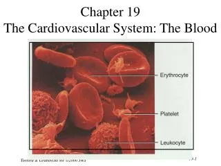
Development of the Driving Pressure in the Human Cardiovascular System 102 Arterial 100 Pressure Normal Resting Pressure Driving the Blood from Left Ventricle to Vena Cava: 102 - 2 = 100 mmHg (mm Hg) 26 7 Mean Circulatory Filling Pressure 7 0 7 6 7 2 Central Venous Pressure (mm Hg) 1 5 0 Normal Resting Cardiac Output Cardiac Output (Q) (Liters / min)
Mean arterial pressures in red The “Closed” Cardiovascular Hemodynamic System LV PO2 = 160 PCO2 = .3 RV LA RA LUNGS AORTA (13) (100) (3) (0) PO2 = 100 PCO2 = 40 9% of blood volume (7) SYSTEMIC ARTERIES Ohms Law: Flow (Q) = upstream pressure – downstream pressure resistance (92) low compliance 13% of blood volume VEINS (CAPACITANCE VESSELS) (20) high compliance 64% of blood volume (40) (2) CAPILLARYBEDS ARTERIOLES PO2 = 40 PCO2 = 46 7% of blood volume Systemic Circulation = 100 mmHg – 0 mmHg = 100 ml / sec = 6 liters / min Flow (Q) 1 mmHg sec / ml
Calculation of Cardiac Output andTotal Peripheral Resistance • Normal Resting Blood Flow ( Q ) = 5 - 6 Liters / min • Recall that: Flow = Pressure • Pressure = Flow X Resistance • Since Resistance = Pressure = Where: V = blood viscosity L = vessel length r = vessel radius Resistance Q 8 V L p r 4 8 V L p r 4
Procedures for Blood Pressure Assessment • Pick the proper cuff for your subject. You can choose from adult and large adult sizes. The cuffs also come in sizes for infants and children. The bladder of the cuff should wrap about 80% of the way around the arm. • With the subjects arm straight, apply moderate pressure around the antecubital space and locate the pulse of the subject on his/her dominant arm. It is usually on the medial side of the antecubital space. Mark this spot with a felt tip pen if necessary. • Place the stethoscope in your ears with the earpieces pointing toward the front of your face and the sounding pad on the subject’s pulse location. Pump the cuff slowly until you hear a thumping sound (signifying that you can, indeed, hear the Korotkoff sounds). Then continue to pump the cuff to about 170 mmHg (or until the sounds have clearly disappeared) and slowly release the valve. Watch the mercury fall and listen for the Korotkoff sounds. Note the points on the mercury column when the sounds occur……… • KOROTKOFF SOUNDS - the sounds heard in the stethoscope marking the different phases of BP • Phase I - First appearance of any type of rhythmic thumping sound (SBP) • Phase II - A murmur or swish • Phase III - Crisper sounds increasing in intensity • Phase IV - Sound becomes muffled (DBP..…???) • Phase V - Sound disappears (DBP..…???)For adults the first sounds mark systolic blood pressure and the point where sound becomes muffled (Phase IV) marks diastolic blood pressure. Listen carefully for the other sounds, but be sure to note Phase I, IV, and V. Phase IV and V may be inseparable at rest but are often widely separated during exercise or in young children. For this reason, the AHA recommends that pressures corresponding to Phases I, IV, and V be recorded. However, the standard and most common method for reporting blood pressure is to report the systolic blood pressure (Phase I) over the diastolic blood pressure (Phase IV or V, whichever comes first).
Lab Assignment for Data Collection #2 Directions: Students should work in groups of 2 with each student serving as both a subject and a data collector. Each student will complete the assignment using his partner’s data. Record all requested information and perform all necessary calculations (SHOW YOUR WORK AND CALCULATIONS). • Resting and orthostatic measurements: Have your subject to lie in a supine position with his/her feet elevated for about 5 minutes. Measure and record the supine systolic and diastolic blood pressure (BP). Next, have the subject stand upright and immediately repeat the BP measurement. (Start pumping up the cuff while the person is still lying down so that your are starting to let the mercury fall just as the person attains an upright position.) Record all values on a data sheet. • Treadmill Assessment: Using one of the treadmills, conduct 3 consecutive stages of exercise for 3 minutes each: 2.0mph @ 10%, 3.2 mph @ 12%, 4 mph @13%. 15 seconds before the end of each stage, take the BP and record it on a data sheet. After completing the last stage, bring the treadmill down to 2.0 mph then stop the belt. Remove the subject from the treadmill and allow them to lie on the patient table for 5 minutes. Take the supine blood pressure. Record all values on a data sheet. • Cycle Assessment. Calculate 3 workloads on the bike that are equivalent to the workloads on the treadmill. Correctly position the subject on the Monarch bike and administer the 3 workloads for 3 minutes each to the subject. Take the subject’s BP 15 seconds before the end of each stage.After completion of the last stage,remove the tension from the belt, take the subject off the bike, and allow them to lie on the patient table for 5 minutes. Take the supine blood pressure. Record all values on a data sheet. Note: treadmill and cycle workloads may be adjusted to accommodate the subject. Just make sure the stages of the treadmill and cycle protocols match in terms of rate of oxygen consumption.
Lab Work-up for Assignment #2 • Using a graphing program (PowerPoint, Excel, etc), plot the SBP, DBP, and MAP [.3 ( SBP - DBP ) + DBP)] for the first supine measurement in section 1, the standing measurement in section 1, the three stages of the treadmill exercise bout in section 2, plus the 5 minute supine recovery. Plot all 3 dependent (y-axis) variables on one graph against the labels “supine”, “standing”, workload 1, workload 2, workload 3, and “recovery” - express all three workloads on the x-axis in METS. • Briefly explain what physiological mechanisms are responsible for differences you found between the resting supine and the standing blood pressures along with the increase in SBP blood pressure that occurs when going from rest to exercise. • Compare the blood pressures during exercise on the treadmill with the corresponding equivalent workloads on the bike. Provide physiologic rationale for any differences noted. • Using a Physician’s Desk Reference (available in the Medical Library or here in our lab) describe in detailhow (by what pharmacological / physiological mechanisms) the following drugs will alter blood pressure at rest. Inderal, Calan, Nitrobid, Accupril, Lasix.