Download

1 / 47
470 likes | 722 Vues
Alterations in Physical Integrity. Types of Wounds. Wound: disruption of normal anatomical structure and FX that results from pathological processes beginning internally or externally to the involved organ(s). (p. 1551). Classification of Wounds. Open vs. Closed. Acquisition. Contamination.

E N D
Types of Wounds Wound: disruption of normal anatomical structure and FX that results from pathological processes beginning internally or externally to the involved organ(s). (p. 1551)
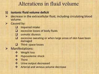
Acute: Wound that proceeds through an orderly and timely reparative process. • Chronic: Wound that fails to proceed through an orderly and timely reparative process. • Superficial: Wound that involves only epidermal layer of skin.
Stages of Wound HealingRegeneration: The process of tissue renewal • Defensive stage (Inflammatory Phase/Reaction) (hemostasis, inflammation, cell migration & epithelialization)
Reconstructive stage (Proliferative Phase/Regeneration) • Filling in of the wound with new connective or granulation tissue • the closing of the top of the wound by epitheliazation.
Maturative stage (Maturation Phase /Remodeling) May take more than a year. Collagen scar continues to reorganize and gain strength for several months. Usu. scar tissue has fewer pigmented cells and has a lighter color than normal skin.
Classification of Wound Healing Primary Intention • Wounds that heal with little tissue loss. • The skin wedges are approximated. • Risk of infection is low. • Healing occurs quickly: drainage stops by day 3 of closure, wound is epitheliazed by day 4, inflammation is present up to day 5, healing edge is present by day 9.
Classification of Wound Healing Secondary Intention Wound edges do not approximate. Wound is left open until it becomes filled by scar tissue. Chance of infection is greater.Inflammatory phase is often chronic Wound filled with granulation tissue (a form of connective tissue that has a more abundant blood supply than collagen. Scarring is greater.
Classification of Wound Healing Tertiary Intention There is a time delay between the time of the injury and the approximation of the wound edges. Attempt by surgeon to allow for effective drainage and cleansing of a clean-contaminated or contaminated wound. Not closed until all evidence of edema and wound debris has been removed. Dressing is used to protect.
Wound Drainage Serous: Clear, watery Sanguineous: Hemorrhagic. Specify color. Serosanguinous: pink to light red in color. Thinner than sanguineous. Purulent: thick drainage that is often yellow-green in color.
Complications of Wound Healing • Hemorrhage • Dehiscence • Evisceration • Infection • Fistulas
Nursing Process for Wound Management Untreated Wounds – basic first aide Treated Wounds – prescribed per M.D. or wound care nurse. Wound Care Protocol
Wound Assessment • Appearance • Drainage (penrose, J-P drain, Hemovac) • Swelling & Induration • Pain • Temperature
Sequential signs of primary wound healing: • Absence of bleeding • Inflammation • Granulation tissue • Scar formation • Reduction in scar size
Lab Data WBC Hgb, Hct BUN, Albumin Wound cultures
MD promotes wound healing RN provides: • Ongoing wound assessment • Aseptic wound care according to MD specifications • Documentation of wound status • Keeps MD apprised of wound status as appro.
To promote healing/prevent complications… • Adequate nutrition • Prevent wound stress/trauma vomiting coughing abdominal distention • Prevent wound infection
Factors Affecting Wound Care • Type of wound • Size • Drainage/exudate • Open vs. closed • Wound location • MD orders • Presence of complications
Drain management • Open vs. closed • Monitor drainage • Universal precautions, aseptic technique
Penrose Drain Open Drainage System
Jackson Pratt Drain Close Drainage system
Hemovacs Drainage Collection Bag (T-tubes) Close Drainage System
Sutures…. Staples…. Hot/cold applications
Pressure ulcerPressure sore, Decubitus Ulcer • Epidermis: Stratum corneum stratum basale • Dermis
Tissue Ischemia: local absence of blood flow/major reduction in blood flow Blanching: Normal red tones of light-skinned client are absent. Does not occur in clients with darkly pigmented skin. Darkly pigmented skin: Skin that remains unchanged (does not blanch) when pressure is applied over a boney prominence – irrespective of the client’s race or ethnicity.
Normal Reactive Hyperemia: Visible effect of localized vasodilatation, the body’s normal response to lack of blood flow to the underlying tissue. Area blanches with fingertip pressure. Lasts less than 1 hour. Abnormal reactive hyperemia: Excessive vasodilatation and induration in response to pressure. The skin appears bright pink to red. Lasts more than 1 hour to 2 weeks after the removal of the pressure. Does not blanch.
Characteristics of Intact Dark Skin that might alert nurses to the potential for pressure ulcers (p. 1546) Color Temperature Touch Appearance
Risk Factors for Skin Breakdown Impaired Sensory input Impaired motor fx Alteration in LOC Orthopedic devices Any equipment
Shearing Force Friction Edema Anemia Cachexia Obesity Infection Impaired peripheral circulation Age (elderly) Nutrition Contributing Factors
Stage I (no skin loss)
Stage I (no skin loss)
Stage II (Shallow crater – involves epidermis and/or dermis)
Stage II Shallow crater – involves epidermis and/or dermis)
Stage III (Full thickness involving damage/necrosis of subc. Tissue. Does not extend down through underlying fascia)