Download
1 / 30
310 likes | 426 Vues
Vascular System. Learning objectives. To describe the components of the vascular system. To understand the structure of arteries, veins and capillaries. To understand what is meant by the term ‘vascular shunt’.

E N D
Vascular System Learning objectives To describe the components of the vascular system. To understand the structure of arteries, veins and capillaries. To understand what is meant by the term ‘vascular shunt’. To be able to describe blood pressure and blood velocity and how these change in response to exercise. To understand how blood pressure is controlled.
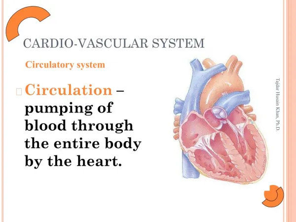
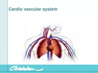
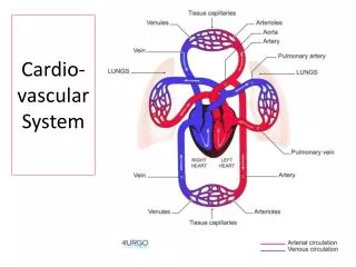
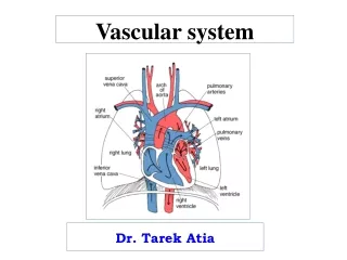
The Vascular System The vascular system is made up of the heart, blood & the blood vessels. Circulatory system introduction VIDEO CLIP.
The Vascular System There are two types of circulation. The pulmonary circulation takes deoxygenated blood from the heart to the lungs and oxygenated blood back to the heart. The systemic circulation carries oxygenated blood to the body from the heart and returns deoxygenated blood from the body to the heart.
The pulmonary circuit The systemic circuit
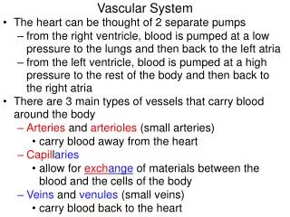
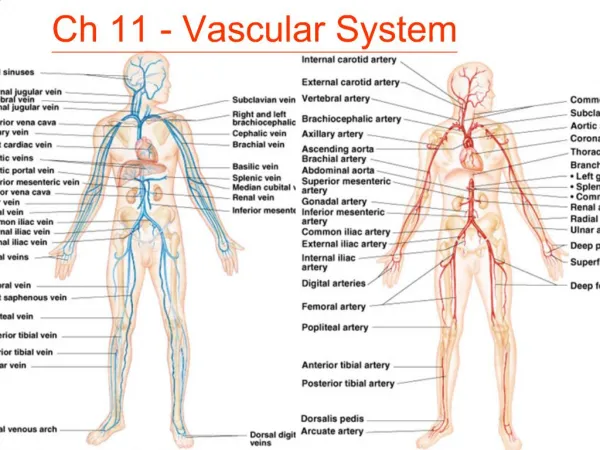
Blood Vessels The vascular network comprises of arteries, arterioles, capillaries, veins and venules. Arteries and arterioles: Arteries are high pressure vessels which carry blood from the heart to the tissues. The largest artery in the body is the aorta which is the main artery leaving the heart. The constant subdivision decreases the diameter of the vessel arteries, which now become arterioles.
Structure of Arteries and Veins Arteries/Viens are composed of three layers of tissue: 1 - an outer fibrous layer — tunica externa 2 - a thick middle layer — tunica media 3 - a thin lining of cells to the inside — tunica interna. The tunica media is comprised of smooth muscle and elastic tissue, which enables the arteries and arterioles to alter their diameter. 3 2 1
Structure of Arteries Arteries will increase the diameter through vasodilation or decrease the diameter through vasoconstriction. It is through vasoconstriction and vasodilation that the vessels can regulate blood pressure and ensure the tissues are receiving sufficient blood particularly during exercise.
Veins and Venules Veins are low pressure vessels which return blood to the heart. The structure is similar to arteries, although they possess less smooth muscle and elastic tissue. Venules are the smallest veins and transport blood away from the capillary bed into the veins. Veins gradually increase in thickness the nearer to the heart they get, until they reach the largest vein in the body, the vena cava, which enters the right atrium of the heart.
Veins and Venules The thinner walls of the veins means blood pools in them. Veins contain pocket valves which close intermittently to prevent back flow of blood. This explains why up to 70% of total blood volume is found in the venous system at any one time.
Capillaries Capillaries are the functional units or the vascular system. They are just thin enough to allow red blood cells to squeeze through their wall. The capillary network is very well developed as they are so small. Large quantities are able to cover the muscle, which ensures efficient exchange of gases.
Veins Recap/Summary information: Before looking at venous return it is important to look at the structure of veins. Veins have thinner walls than arteries. Veins also have valves.
Venous Return Mechanism The heart can only pump out as much blood as it receives, so cardiac output is dependent on venous return. A rapid increase in venous return enables a significant increase in stroke volume and therefore cardiac output. Venous return is overcome in a number of ways: Skeletal Pump Inspiration/Expiration – Respiratory pump Valves Smooth Muscle
Skeletal Pump The muscles surrounding the veins expand and contract, pressing on veins and causing a pumping effect. This muscle action is particularly important in maintaining venous return during exercise. It is referred to as the skeletal pump.
Respiratory Pump Muscles around the thoracic and abdominal regions cause changes in pressure. Change in pressure allow the veins in this region to compress, causing blood to be ‘sucked’ through them.
Valves The blood in the veins can only move towards the heart; It cannot fall back to where it came from. This is because at regular intervals there are semi lunar pocket valves situated in large veins. The prevent blood from flowing away from the heart.
Blood flow changes during exercise Look at the changes in blood redistribution in the diagram shown. What do you notice? Why does this shunt take place?
Vascular Shunt During exercise, blood flow to the skeletal muscles increases to meet the increase in oxygen demand. This redirection of blood flow to the areas where it is most needed is known as a vascular shunt.
Vascular Shunt Sports performers should not eat within an hour of competition. Eating results in blood being redirected to the stomach to aid digestion. This would affect performance because less oxygen would be available for the muscles.
Redistribution of Blood Flow The redistribution of blood is controlled primarily by the vasoconstriction and vasodilation of blood vessels. Decreases blood flow through narrowing Increases blood flow through widening
Vasodilation/Vasoconstriction It reacts to chemical changes of the local tissues. For example, vasodilation will occur when arterioles sense a decrease in oxygen concentration or an increase in acidity due to higher CO2 and lactic acid concentrations. Sympathetic nerves in the TUNICA MEDIA also play a major role in redistributing blood from one area of the body to another.
Vasodilation/Vasoconstriction Further structures which aid blood redistribution are pre-capillary sphincters. These are ring shaped muscles which lie at the opening of capillaries and control blood flow into the capillary bed. When the sphincter contracts, it restricts blood flow through the capillary, and deprives tissues of oxygen. When it relaxes, it increases blood flow to the capillary bed.
T Myoglobin In the muscle, oxygen is stored by myoglobin. This has a high affinity for oxygen and stores the oxygen until it can be transported from the capillaries to the mitochondria. The mitochondria are the sites in the muscle where aerobic respiration takes place.
Blood Pressure DEFINITION: Blood pressure is the force exerted by the blood against the walls of the blood vessels. It is necessary to maintain blood flow though the circulatory system • It is determined by two main factors – • Cardiac Output – the volume of blood flowing into the system from the left ventricle. • Resistance to flow – the impedance offered by the blood vessels to the blood flow.
Blood Pressure Blood pressure = cardiac output x resistance Therefore, blood pressure increases when either cardiac output or resistance increases. Blood pressure in the arteries also increases and decrease in a pattern which corresponds to the cardiac cycle during ventricular systole, when blood is pumped into the aorta and lowest during ventricular diastole. Systole Diastole
Blood Pressure Measurement BP is usually measured at the brachial artery using a sphygmomanometer, and is recorded as mmHg. SYSTOLE pressure (TOP FIGURE) is experienced when the heart pumps blood into the system DIASTOLIC pressure (BOTTOM FIGURE) is recorded when the heart is relaxing and filling with blood. Typical Reading = 120mmHg 80mmHg
Blood Pressure and Exercise Blood Pressure changes during exercise. During aerobic exercise, the systolic pressure increases as a result of increased cardiac output, while diastolic pressure remains constant. During anaerobic exercise both systolic and diastolic pressure rise significantly due to increased resistance of the blood vessels.
Blood Velocity Speed / velocity of blood flow changes as blood travels through the systemic circulation. TASK: Discuss where it might it be at its fastest? It is fasted in the aorta and other large arteries. Slowest in the capillaries and veins. BUT picks up speed in the veins leading up to the heart. 50403020100 Velocity of blodd flow (cm/s) Venae Cavae Veins Aorta Venules Arteries Arterioles Capillaries
Blood Velocity 500040003000200010000 Velocity is inversely (THE OPPOSITE) related to cross-sectional area of the blood vessels to be filled. Blood flows fastest where the total cross-sectional area is the least. Total area (cm2) of vascular bed 50403020100 Velocity of blodd flow (cm/s) Venae Cavae Veins Aorta Venules Arteries Arterioles Capillaries
Control of blood pressure The vasomotor centre controls blood pressure. Baroreceptors located in the aortic and carotid arteries detect increases and decreases in blood pressure and send an impulse to the vasomotor centre located in the medulla oblongata .
Plenary Exam Style Question. 1. Describe the process of vascular shunt and why does the body respond in this way? (4 marks) 2. What is the venous return mechanism and what 4 mechanisms affect its efficiency (4 marks)