Download
1 / 34
350 likes | 850 Vues
Cardio-vascular system. Outline. 1- Overview 2- Path of blood through the heart and vasculature 3- Anatomy of the heart 4- Electrical activity of the heart 5- The cardiac cycle 6- Cardiac output and its control. Outline. 1- Overview 2- Path of blood through the heart and vasculature

E N D
Outline • 1- Overview • 2- Path of blood through the heart and vasculature • 3- Anatomy of the heart • 4- Electrical activity of the heart • 5- The cardiac cycle • 6- Cardiac output and its control
Outline • 1- Overview • 2- Path of blood through the heart and vasculature • 3- Anatomy of the heart • 4- Electrical activity of the heart • 5- The cardiac cycle • 6- Cardiac output and its control
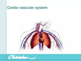
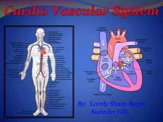
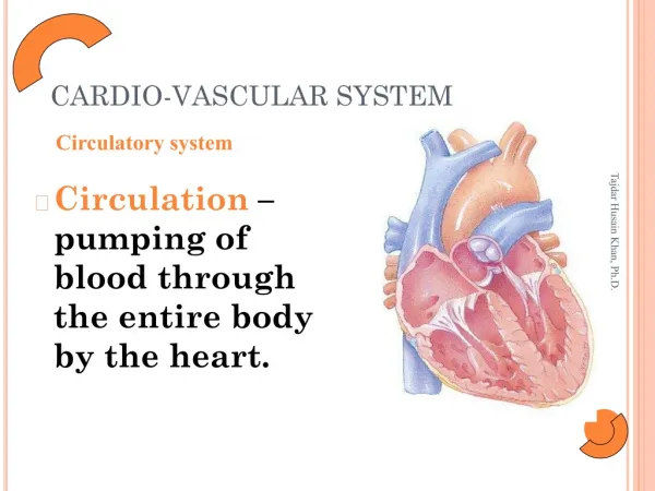
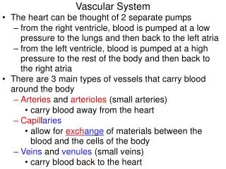
Roles: - Pumps blood throughout the body vasculature - Endocrine function Components - Heart - Blood vessels - Blood Overview
Outline • 1- Overview • 2- Path of blood through the heart and vasculature • 3- Anatomy of the heart • 4- Electrical activity of the heart • 5- The cardiac cycle • 6- Cardiac output and its control
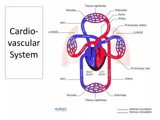
Parallel flow of blood to various organs -- allows for fully oxygenated blood to reach each organ -- allows for independent regulation Exception: portal circulation (1 capillary bed to another) -- hypothalamus-pituitary gland portal system -- hepatic portal system Circulation Figure 13.3
Applications • What is the consequence of the blood clot (a thrombus) located in the right saphenous vein becoming loose ( an embolus)? • What is the consequence of the blood clot (a thrombus) located in the right atrium becoming loose ( an embolus)? • What is the consequence of the blood clot (a thrombus) located in the left atrium becoming loose ( an embolus)? • What is the consequence of the blood clot (a thrombus) located in the left saphenous vein becoming loose ( an embolus)? • What is the consequence of the blood clot (a thrombus) located in the right femoral artery becoming loose ( an embolus)?
Located in mediastinum Surrounded by the pericardium - outer fibrous pericardium - inner serous pericardium: - parietal pericardium - visceral pericardium - in between: pericardial cavity small amount of pericardial fluid (prevent friction) Application: cardiac tamponade The heart
Outline • 1- Overview • 2- Path of blood through the heart and vasculature • 3- Anatomy of the heart • 4- Electrical activity of the heart • 5- The cardiac cycle • 6- Cardiac output and its control
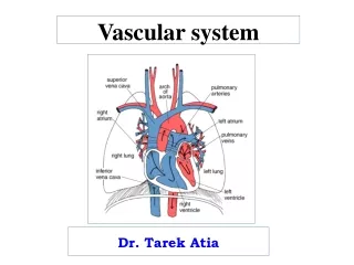
The heart: review Figure 13.1
Blood flow in the heart Figure 13.6
What is angina? What is a myocardial infarction? Coronary circulation Figure 13.4
Outline • 1- Overview • 2- Path of blood through the heart and vasculature • 3- Anatomy of the heart • 4- Electrical activity of the heart • 5- The cardiac cycle • 6- Cardiac output and its control
Two types of fibers: - contractile fibers (cardiac muscle fibers) - self-depolarizing fibers in the sino-atrial (S/A) node (pace-maker fibers autorhythmicity) Conduction system of the heart Figure 13.10
Recording of the electrical activity of the heart by electrodes applied on the skin ECG wave patterns vary with the location of the electrodes Electrocardiogram
P wave: S/A node is firing P-Q interval: time it takes for the electrical impulse to travel from the S/A node to the atrio/ventricular (A/V) node QRS wave: the electrical impulses spread through the bundle of His, bundle branches and Purkinje fibers in the ventricles T wave: ventricular repolarization Q-T interval: corresponds to ventricular contraction (=systole) T-Q interval: ventricular diastole R-R interval: time between heartbeats ECG
Other properties of the conduction system • S/A node: 60-100 beats/min (sinus rhythm = normal rhythm) • If S/A node is non functional: the A/V node takes over 40-60 beats/min • If both S/A and A/V nodes are shot, ventricular electrical activity takes over > 40 beats/min
Applications • What is atrial fibrillation? • “ “ ventricular fibrillation? • What is the difference between tetanus and fibrillation? • Atrial and ventricular fibrillations: consequences from each type of abnormal rhythms
Outline • 1- Overview • 2- Path of blood through the heart and vasculature • 3- Anatomy of the heart • 4- Electrical activity of the heart • 5- The cardiac cycle • 6- Cardiac output and its control
Cardiac cycle Figure 13.18
Systole: contraction of heart chambers but mostly ventricles Diastole: ventricular relaxation (atria have a minimal effects) Ventricular filling: during diastole, P wave Ventricular contraction: at first, semi-lunar valves are closed blood cannot flow out and pressure increase in ventricle isovolumetric contraction Ventricular ejection: The pressure against the valves is strong enough to open them the blood flows out When the ventricles stop contracting, the pressure falls isovolumetric relaxation pressure falls even more semi-lunar valves close and A/V valves open Cardiac cycle
End-diastolic volume = EDV = volume of blood present at the end of diastole in the ventricles End-systolic volume = ESV = volume of blood present at the end of systole Stroke volume = SV: amount of blood ejected by the ventricles = EDV-ESV Ejection fraction = EF = SV/EDV Note: EF gives a measure of cardiac muscle efficiency Cardiac cycle
What can cause a low ejection fraction? • What are the consequences of a low ejection fraction?
Outline • 1- Overview • 2- Path of blood through the heart and vasculature • 3- Anatomy of the heart • 4- Electrical activity of the heart • 5- The cardiac cycle • 6- Cardiac output and its control
Cardiac output = CO • Cardiac output = volume of blood pumped out by the heart per minute • CO = SV x HR (CO must adapt to body needs) • Control of CO: ** control of SV: - Intrinsic control - Extrinsic control ** control of HR: - Extrinsic control -- Autonomic input -- Hormonal control
SV: a function of 1. Ventricular contractility: a function ventricular health and stretch 2. EDV: ventricular refill is a function of the blood pressure in the central veins (central venous pressure) end- diastolic pressure = preload 3. ESV: a function of afterload = pressure against blood flow out of the heart – determined by aortic blood pressure Control of the stroke volume
Cardiac output = CO • Cardiac output = volume of blood pumped out by the heart per minute • CO = SV x HR (CO must adapt to body needs) • Control of CO: ** control of SV: - Intrinsic control - Extrinsic control ** control of HR: - Extrinsic control -- Autonomic input -- Hormonal control
Intrinsic control - Starling law of the heart: The heart automatically adjust its output to match its input - Property of the cardiac muscle: the more it is stretched, the stronger it contracts (up to a limit) (in other word, what ever comes in, goes out) What would happen if this law is not respected? Control of the stroke volume
Extrinsic control Neural control: -- the sympathetic NS has axonal extension over the entire ventricles β receptors binding to NE stronger contraction -- no parasympathetic axonal extension no direct action on ventricular wall Hormonal control -- Epinephrine from adrenal medulla has the same effect as NE from sympathetic nerve endings increased force of contraction Control of the stroke volume
Cardiac output = CO • Cardiac output = volume of blood pumped out by the heart per minute • CO = SV x HR (CO must adapt to body needs) • Control of CO: ** control of SV: - Intrinsic control - Extrinsic control ** control of HR: - Extrinsic control -- Autonomic input -- Hormonal control
S/A node fires automatically 60-100/times per minute. Its activity is modulated by the following factors: Extrinsic control only -- Autonomic NS * action on the S/A node mainly * NE increases HR * Ach decreases HR -- Hormonal control * Epinephrine from the adrenal gland increases HR -- drugs and ions (K+, Ca++, digoxin and others) Control of heart rate (HR)
The cardiac center in the medulla oblongata controls the HR (or S/A node). It receives information from the body through various receptors Aortic and carotid bodies monitor blood O2 and send the info. to the cardiac center (↓O2↑HR) CO2 and pH receptors in the hypothalamus also send info. to the cardiac center (under normal conditions, they have more influence on HR then O2 receptors (↑CO2 or ↓pH ↑HR) Body temperature (↑Temp ↑HR) Factors influencing HR
Applications • Jimmy has an abnormal HR at 144b/min. He has been admitted and is on medication so his HR reverts to a sinus rhythm (when the S/A node is in control). The next day, his HR is unchanged. • Roger also has an abnormal HR at 131b/min. He has been admitted and is on medication so his HR reverts to a sinus rhythm (when the S/A node is in control). The next day, his HR is unchanged. • Marian has been admitted during the night. She has a sinus rhythm (driven by S/A node) at 125 b/min. • Carlie has an abnormal HR at 55 b/min. He has been admitted and is on medication so his HR reverts to a sinus rhythm (when the S/A node is in control). The next day, his HR is unchanged. • Which of these 4 patients would you go see first? Why? • Hint: how is the HR regulated?
Applications • Jimmy has been on medication for an abnormal HR at 144 b/min. On a cardiac monitor, you see his heart rate jumping to 190 b/min. Which consequences do you expect?


![CARDIO-VASCULAR SYSTEM [CVS] FUNCTIONAL ANATOMY OF HEART](https://cdn1.slideserve.com/1739818/cardio-vascular-system-cvs-functional-anatomy-of-heart-dt.jpg)