Download
1 / 219
2.25k likes | 2.32k Vues
Cardio vascular system. CARDIO VASCULAR SYSTEM. INDEX 1) INTRODUCTION OF CARDIO VASCULAR SYSYTEM 2) THE HEART 3) PROPERTIES OF CARDIAC MUSCLES 4) CONDUCTIVE SYSTEM 5) CARDIAC CYCLE 6) HEART SOUND 7) HEART RATE 8) ELECTRO CARDIO GRAM 9) CARDIAC OUTPUT

E N D
INDEX 1) INTRODUCTION OF CARDIO VASCULAR SYSYTEM 2) THE HEART 3) PROPERTIES OF CARDIAC MUSCLES 4) CONDUCTIVE SYSTEM 5) CARDIAC CYCLE 6) HEART SOUND 7) HEART RATE 8) ELECTRO CARDIO GRAM 9) CARDIAC OUTPUT 10) REGULATION OF CARDIAC FUNCTION 11) BLOOD PRESSURE 12) APPLIED PHYSIOLOGY OF THE HEART 13) SHOCK
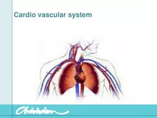
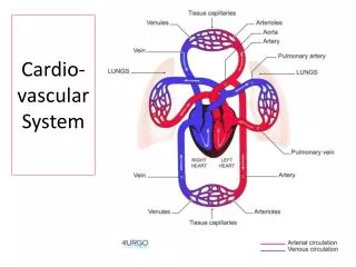
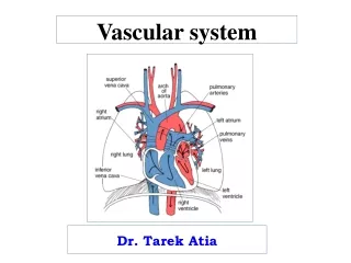
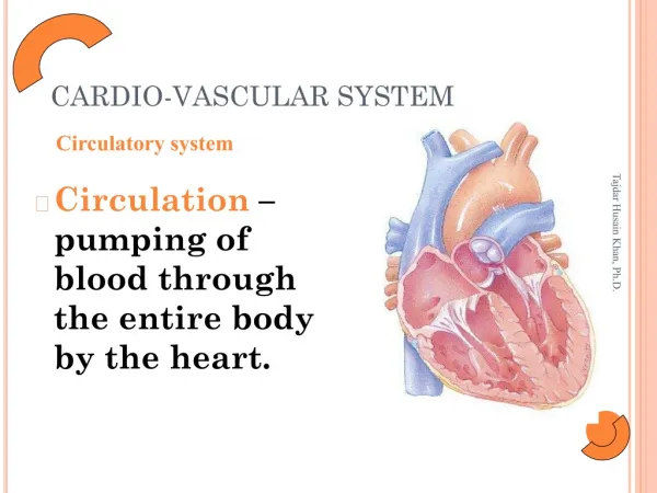
Cardio Vascular System Introduction:- Cardio Vascular System is a system of circulation. Cardio vascular system includes. Heart – It is a pumping organ of the body. Arteries - If carries oxygenated blood to all parts of the body. Capillary – Through it water and other small molecule substance passes. Veins – It carries deoxygenated blood from all parts of the body.
The cardio – Vascular system is divided, for descriptive purpose, into two main parts. • The circulatory system, consisting of the heart, which acts as a pump, and the blood vessels through which the blood circulates. • The lymphatic system, consisting of lymph nodes and lymph vessels , though which colourless lymph flows .
CIRCULATORY SYSTEM:- The circulatory system consist of blood vessels namely aorta, arteries, arterioles, capillaries, venules ,veins and vena cava.
DIVISIONS OF CIRCULATORY SYSTEM • SYSTEMIC CIRCULATION :- This is also known as greater circulation. The blood , which is pumped from left ventricle passes through a series of blood vessels of arterial free or arterial system and reaches the tissues. The blood vessels of the arterial are the aorta , larger arteries, smaller arteries and arterioles . The arterials branch into the capillaries. The capillaries are responsible for exchange of various substances between blood and the tissues.
2. PULMONARY CIRCULATION:- This is also called lesser circulation blood is pumped from right ventricle to lungs through pulmonary artery . The exchange of gases occurs between blood and alveoli of the lungs though pulmonary capillary membrane . The oxygenated blood returns to left atrium through the pulmonary veins Thus, the left side of the heart contains oxygenated as arterial blood and the right side of the heart contains the venous blood.
FUNCTIONS OF CARDIO –VASCULAR SYSTEM The primary function of the cardio vascular System is to provide an adequate supply to all cells of the body, of materials needed for their proper function and that carries away the waste products and that carries away the waste products of their metabolism It is a well organized transport system of the body by which the blood being circulation within a closed system under different pressure gradients created by the pumping mechanism where heart acts as the central pump.
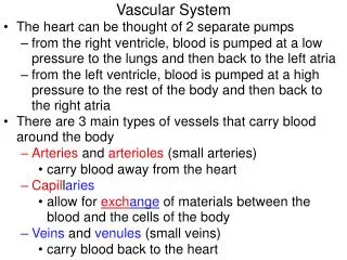
HISTOLOGY OF CARIDO –VASCULAR SYSTEM:- The aorta first , breaks up into a number of big arteries , each one of which divides into a number of smaller arteries. Each small arteries give rise to a bunch of arterioles. Blood then flows from arteriole into a meta arteriole collect into venules , venules into vein and ultimately come back to the heart through the two vena cava. ARTERIES AND ARTERIOLES:- These are the blood vessels that transport blood away from the heart . Their walls consist of three of tissue.
Tunica adventitia or outer layer of fibrous tissue • Tunica media or middle layer of smooth muscle and elastic tissue • Tunica intima or inner lining of squamous epithelium called endothelium VEINS AND VENULES Veins are blood vessels that return blood at low pressure to the heart . The walls of the veins are thinner than those of arteries but have the same three layer of tissue. They are thinner because there is less muscle and elastic tissue in the tunica media, because veins carry blood at lower pressure than arteries when cut, the veins collapse while the thicker –walled arteries remain open .
when an artery is cut blood spurts at high pressure while a slower, steady flow of blood escapes from a vein .veins posses the values which prevents the backflows of blood. The smallest veins are called venules. CAPILLARIES AND SINUSOIDS:- The smallest arterioles break up into number of minute vessels called capillaries. Capillary walls consist of a single layer of endothelial cell sitting on a very thin basement membrane , through which water and small-molecule substance can pass.
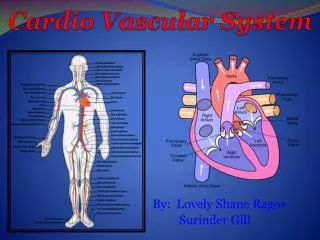
HEART Anatomy of the Heart POSITION:- The heart lies in the thoracic cavity. In the mediastinum. It lies obliquely , a little more to the left than the right, and presents a base above, and an apex below. The apex is about 9cm to the left of the midline at the level of the 5thintercoastal space, i.e a little below the nipple and slightly nearer the midline. The base extends to the level of the 2nd rib. Organs Associated With The Heart Inferiorly – the apex rests on the central tendon of the diaphragm
Superiorly – the great blood vessels i.e. the aorta, superior vena cava, pulmonary artery and pulmonary veins. Posteriorly - The oesphagus trachea left and right bronchus descending aorta inferior vena cava and therocic vertebrae Laterally- the lungs, the left lung over laps the left side of the heart Anteriorly- the sternum ribs and intercostals muscles. STRUCTURE • The heart is composed of three layers of tissue pericardium , myocardium and endocardium.
APPLIED– CARDIOPALMONARY RESUSCITATION • Because the heart lies between two ridge structure vertebral column and the sternum –external pressure on the chest can be used to force blood out of the heart and into the circulation. In cases in whichthe heart suddenly stops beating – cardiopulmonary resuscitation (CPR) – properly cardiac compression performed with artificial ventilation of the lungs via mouth to mouth respiration saves lives. PERICARDIUM:- • The pericardium is made up of two sacs. The outer sac consists of fibrous tissues and the inner of a continuous double layer of the serous membrane, the parietal pericardium, lines the fibrous sac.
The line layer, the visceral pericardium or pericardium, which is in continuous with the parietal pericardium, is adherent continuous with the parietal pericardium is adherent to the heart muscle flattened epithelial cells. Between the parietal and visceral layer of the serous pericardium is a thin film of serous fluid . This slippery secretion of the pericardial cells, is known as pericardial fluid reduces friction between the layer of the serous pericardium ad the heart moves. The space that contains the fluid is called the pericardial cavity.
. APPLIED PERICARDITIS Inflammation of the pericardium is called pericarditis. As a result of inflammation to the pericardium there is chest pain that may extend to the left shoulder and down the left arm. Acute pericarditis usually lasts for about one week and is treated with drugs that reduce inflammation and pain such as ibuprofen or aspirin.
MYOCARDIUM The myocardium is composed of specialised cardiac muscle found only in the heart. It is mot under voluntary control but like skeletal muscle, cross – stripes are seen on microscopic examination. The myocardium is thickest at the apex and thins out towards the base. This reflects the amount of work each chamber contributes to the pumping of blood. It is thickest in the left ventricle, which has the greatest workload. APPLIED MYOCARDITIS: Myocarditis is an inflammation of the myocardium that usually occurs as a complication
of a viral infection, rheumatic fever or exposure to radiation or certain chemicals or medication. Myocarditis often has no symptoms. However, if they do occur they may include fever, fatigue, vague chest pain irregular or rapid heart beat, joint pain and recovery occurs within two weeks. Severe cases can lead to cardiac failure and death ENDOCARDIUM The lines the chambers and valves of the heart. It is a thin smooth, glistening membrane that permits smooth flow of blood inside the heart. It consists of flattened epithelial living the blood vessels.
ENDOCARDITIS It refers to an inflammation of the endocardium and typically involves the heart valves. Most cases are caused by bacteria signs and symptoms of endocarditis include fever, heart murmur, irregular or rapid heart beat, fatigue, loss of appetite night sweats and chills. CHAMBERS OF THE HEART The heart has four chambers. The two superior chambers are the atria and two inferior chambers are the ventricles . On the anterior surface of each atrium is a wrinkled pouch like structure called an auricle. RIGHT ATRIUM The right atrium receives blood from three veins the superior vena cava, the inferior vena
cava and coronary sinus. The anterior and posterior walls of the right atrium are very different. The posterior wall is smooth the anterior wall is rough due to the presence of muscular ridge called pectinate muscles which also extend into the auricle. Between the right atrium and left atrium there is a thin partition called the interarterial septum. Blood passes from the right atrium into the right ventricle through a valve that is called the tricuspid valve because it consists of three leaflets or cusps. It is also called the right atrioventricular valve.
RIGHT VENTRICLE The right ventricle forms most of the anterior surface of the heart. The cups of the tricuspid valve present in right ventricle are connected to tendon like cords the chordae tendinae, which in turn are connected to cone-shaped trabeculae carneae called papillary muscles. The right ventricle is separated from the left ventricle by a partition called the inventricular septum. Blood passes from the right ventricle through the pulmonary value into a large artery called the pulmonary trunk which divides into right and left pulmonary arteries.
. LEFT ATRIUM The left atrium forms most of the base of the heart. It receives blood from the lungs through the four pulmonary veins. Blood passes from the left atrium into the left ventricle through the bicuspid valve which as its name implies it has two cusps. It is also called the left atrioventricular valve. LEFT VENTRICLE The left ventricle forms the open of the heart. Like the right ventricle, left ventricle contains trabeculae corneal and has chordae tendinae. Blood passes from the left ventricle through the aortic vaqlue into the ascending aorta.
PROPERTIES OF CARDIAC MUSCLES • Rythmicity • Conductivity • Excitability • Contractility • All or non Response • Staircase Phenomenon • Refractory Period • Tone
1. RHYTHMICITY:- The ability of a tissue to produce its own impulses regularly is called the rhythmicity. This inherent rhythmicity property is present throughout the cardiac muscles as evident from the electrophysiological studies of the single fibre from the atrium or ventricle, S.A Node, A.V Node, atrial muscle purkinje fibre and also from the ventricular muscle fibre. The rate of rhythmicity:- In S.A Node – 70-80 Per Minute In A.V Node – 40-60 Per Minute In Ventricle is – 20-40 Per Minute Due to the higher rhythmically property of the S.A node, it controls the rest of cardiac muscle and thus the heart
beat at the rhythm of the S.A Node. When the S.A node fails , the A.V node takes the change and if it fails the atrium and afterwards ventricle take the change of maintaining heart beat. Cardiac rhythm is altered following stimulation of vagi or sympathetic nerve supplying the heart. 2 CONDUCTIVITY:- Conduction of impulse produced by S.A node are transmitted to the cardiac muscle. The impulse originated at the S.A node spread over the artria and reaches A.V node through the internodal fibres. All these fibres converge towards the A.V node and interdigitate with fibres of AV node.
The A.V transmits the impulse through out the bundle of his and its branches conduction in the bundle of the his and purkinje fibres is 1m/sec. 3 EXCITABILITY AND CONTRACTILITY:- The cardiac muscle is excited by adequate stimuli and response by contraction, the ability of tissue to give response to stimulus is called excitability, Ability of tissue to shorten in contraction after receiving a stimulus is called contractality.The contractile unit of cardiac muscles is myofibril which contain actin and myosin protein units.During contraction actin and myosin are associated in presence of ATP and thus the fibre is shortened but during rest these are dissociated again.
ALL OR NONE RESPONSE:- If a quiescent heart muscle is stimulated at widely spaced electrical shock of increasing strength is reached but there was no such increasing intensities of stimulation. STAIRCASE PHENOMENON:- In a stannius preparation if the ventricular muscle stimulated with inducted current first few contractions gradually increase in rise and them it becomes study this is known as staircase phenomenon are dissociated again.
REFRACTORY PERIOD:- This is the period in which the muscle does not show any response to a stimulus. The refractory period of the heart is long and can be divided into three parts:- • Absolute refractory Period • Relative refractory Period • Supernormal refractory Period ABSOLUTE REFACTORY PERIOD:- This period extends throughout the whole period of contraction. Absolute refractory period during which the muscles does not show any response at all whatever may be the strength of the stimulus.
RELATIVE REFRACTORY PERIOD:- The relative refractory period is the period during which the muscle shows response to a stimulus. SUPERNORMAL PERIOD:- This period is limited from the point of terminal of repolarisation to the beginning of slow diastolic repolarisation phase. The refractory period is longest in the A.V node, intermediate in the ventricles and least in the atria. TONE:- Heart muscle possess tone. This tone is independent of nerves and can be adjusted in this way,
it can maintain a fairly constant tension upon its varying contents. • The smallest fibres with least glycogen at the nodes. • The broader fibres with more glycogen in the ventricles. • The still border fibres with more glycogen in the atria. • The broadest fibres with abundant glycogen in the purkinjefibres.
SPECIAL FUNCTIONAL TISSUES OF THE HEART OR PACE MAKER TISSUE OF THE HEART OR CONDUCTING SYSTEM OF THE HEART. INTRODUCTION:- Cardiac muscles consists essentially of certain specialised structure which responsible for initiation and transmission of cardiac impulse at higher rate than the rest of the muscle. Those specialised cardiac tissues operates such mechanisms are collectively known as the junctional tissue of the heart. They comprises of the following structure.
Sino-artrial (S.A.) node Atrioventricular A.V. node Bundle of his atrioventricular bundle. The right and left branches of the bundle ending in the Purkinje fibres
HISTOLOGY:- cardiac muscles fibres are separated from each other by the connective tissue endomysium along with blood vessels and lymphatics. The cardiac muscle fibre are not made up of one straight simple cylinder but they have got short cylindrical branches in all directions (in any dimensions).
These branches are coming in contact with that of the adjacent fibres ultimately forming a three dimensional network. Under light microscope these networks appear as syncytium which was also supported as the property of the cardiac muscle that if it contacts it will contact as whole. The sino atrial and atrioventricular nodes and bundle of this are composed of specialised cardiac tissue and contain high amount of glycogen. These have got more sarcoplasm than the rest of the cardiac muscles fibres.
PURKINJE FIBRES:-also contain high amount of glycogen in their sarcoplasm. The atrial muscle fibre is connected with the ventricular muscle fibre only through the budle of his because of fibrous tissue ring keeps the atrial muscle separated from the ventricular muscle. Damage of bundle of his cause dissociation of atrial and ventricular rhythm. SINU ATRIAL NODE(SAN). i) Location:- on the posterior aspect of heart at junction of the superior vena cava (SVC) with right atrium (RA). (i.e. free border of the RA appendix)
iv) Innervation:- it develops from structure on the right of the embryo that is why in adults SAN is innervated by vagus nerves it also receive sympathetic nerve. Fibres predominately of right side from the cervical sympathetic ganglia via the cardiac nerves. 2) Atrio ventricular node (AVN) Location:- posteriorly on the right side of the interatrial septum near the opening of coronary sinus. Structure :- Save as that of SAN. Innervation:- it is a left sided structure of the embryo.Therefore in adults it is innervated.
ii) Dimension:- length-15mm, width 2mm and thickness- 1mm. iii) Structure:- more embryonal in character ie cell outline ill defined highly vascular consist of thin elongated muscle fibres reach in glycogen and mitochondria fusiform in shape with longitudinal striation. These are called P cell or pace maker sales, these fibres normally generated and discharge impulses more rapidly then any other pacemaker tissue and their rate of discharge determines the rate at which the heart beats.
THE CONDUCTION SYSTEM An inherent and rythmical electrical activity is the reason for the hearts continuous beating. The source of this electrical activity is a network of specialized cardiac muscle fibres. These cells are called autorhythmic fibres because they are self excitable. Autorhythmic fibres repeatedly generate action potentials that trigger heart contractions Autorhythmic fibres continuous to stimulate a heart to beat even after it is removed from the body for example to be transplanted into another person although all of its nerves have been cut
By left vagus nerve also receive sympathetic nerve supply primarily from left side Modified atrial muscle fibres from the region of coronary sinus collection wise and unite with the AVN called inter nodal atrial pathways. They conduct impulses from SAN to the AVN by narrowing themselves in 3 bundles Anterior internodal tract of Bachmann. It can also conduct impulses from San directly to LA Middle internodal tract of Wenckebach and Posterior internodal tract of THOREL
3) Atrio ventricular bundle or the bundle of his It takes origin from AVN and then runs upwards to the posterior margin of the membranous interventricular septum and then forwards below it, ensheathed and isolated in a canal. At the anterior part of the membranous inter ventricular septum the bundle divides into a right & a left branch The left branch pierces the membrane and then lies on the upper border of the muscular septum divided into an anterior fascicle and a posterior fascicle The right branch passes down the right side of.
the septum. iii) Both branches divide repeatedly to from a network of fibres lying subendocardially in the ventricles. 4) Purkinje fibres Takes origin from terminal divisions of right and left branch of the bundle of this penetrate the ventricular wall. These fibres are some what large and thicker than the cardiac muscle fibre Length:-10-46 um and diameter 70-80 um with indistinct cell outlines and granular central cytoplasm containing several nuclei the
During embryonic development about 1% of the cardiac muscle fibres become autorhythmicfibres which have two important functions:- They act as a pacemaker setting the rhythm of electrical excitation that causes contraction of the heart. They form the conduction system a network of a specialized cardiac muscle fibres that provide a path for e cycle of cardiac excitation to progress through the heart. The conduction system ensure that cardiac chambers become stimulated to contract in a coordinate in a manner which makes the heart and effective pump.


![CARDIO-VASCULAR SYSTEM [CVS] FUNCTIONAL ANATOMY OF HEART](https://cdn1.slideserve.com/1739818/cardio-vascular-system-cvs-functional-anatomy-of-heart-dt.jpg)