Download
1 / 20
200 likes | 533 Vues
gram-positive spore-forming bacilli SPP. Bacillus. Prepared by Assit.Prof.Dr . Najdat B.Mahdi. Introduction.

E N D
gram-positive spore-forming bacilliSPP.Bacillus Prepared by Assit.Prof.Dr. NajdatB.Mahdi
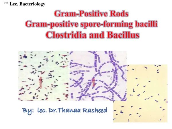
Introduction • The gram-positive spore-forming bacilli are the Bacillus and Clostridium species. These bacilli are ubiquitous, and because they form spores they can survive in the environment for many years. Bacillus species are aerobes, whereas clostridia are anaerobes.
Bacillus Species • The genus bacillus includes large aerobic, gram-positive rods occurring in chains. Most members of this genus are saprophytic organisms prevalent in soil, water, and air and on vegetation, such as Bacillus cereus and Bacillus subtilis. Some are insect pathogens. B cereus can grow in foods and produce an enterotoxin or an emetic toxin and cause food poisoning. Such organisms may occasionally produce disease in immuno- compromised humans (eg, meningitis, endocarditis, endophthalmitis, conjunctivitis, or acute gastroenteritis). B anthracis, which causes anthrax, is the principal pathogen of the genus.
Morphology &Identification • Typical Organisms • The typical cells, measuring 1 x 3–4 µm, have square ends and are arranged in long chains; spores are located in the center of the non-motile bacilli • Culture • Colonies of B anthracis are round and have a "cut glass" appearance in transmitted light. Hemolysis is uncommon with B anthracis but common with the saprophytic bacilli. Gelatin is liquefied, and growth in gelatin stabs resembles an inverted fir tree
Growth Characteristics • The saprophytic bacilli utilize simple sources of nitrogen and carbon for energy and growth. The spores are resistant to environmental changes, with stand dry heat and certain chemical disinfectants for moderate periods, and persist for years in dry earth. Animal products contaminated with anthrax spores (eg, hides, bristles, hair, wool, bone) can be sterilized by autoclaving
Bacillus anthracis • Anthrax is primarily a disease of herbivores—goats, sheep, cattle, horses, etc; other animals (eg, rats) are relatively resistant to the infection
In humans, the infection is usually acquired by the entry of spores through injured skin ( cutaneous anthrax) or rarely the mucous membranes (gastrointestinal anthrax), or by inhalation of spores into the lung (inhalation anthrax). • In inhalation anthrax (woolsorters’ disease), the spores from the dust of wool, hair, or hides are inhaled; phagocytosedin the lungs; and transported by the lymphatic drainage to the mediastinallymph nodes, where germination occurs. This is followed by toxin production and the development of hemorrhagic mediastinitisand sepsis, which are usually rapidly fatel
. In animals, the portal of entry is the mouth and the gastrointestinal tract. Spores from contaminated soil find easy access when ingested with spiny or irritating vegetation. The spores germinate in the tissue at the site of entry, and growth of the vegetative organisms results in formation of a gelatinous edema and congestion. Bacilli spread via lymphatics to the bloodstream, and they multiply freely in the blood and tissues shortly before and after the animal's deaththe number of organisms in the blood • exceeds 107/mL just before death.. • B anthracis that does not produce a capsule is not virulent and does not induce anthrax in test animals. The poly-D- glutamic acid capsule is anti phagocytic. The capsule gene is on a plasmid
Anthrax toxin is made up of three proteins: protective antigen (PA), edema factor (EF), and lethal factor (LF). PA binds to specific cell receptors, and following proteolytic activation it forms a membrane channel that mediates entry of EF and LF into the cell. EF is an adenylylcyclase; with PA it forms a toxin known as edema toxin. LF plus PA form lethal toxin, which is a major virulence factor and cause of death in infected animals. When injected into laboratory animals (eg, rats) the lethal toxin can quickly kill the animals. The anthrax toxin genes are on another plasmid.
In humans, approximately 95% of cases are cutaneous anthrax and 5% are inhalation • Gastrointestinal anthrax is very rare; • The incubation period in inhalation anthrax may be as long as 6 weeks
Diagnostic Laboratory Tests • Specimens to be examined are fluid or pus from a local lesion, blood, and sputum. Stained smears from the local lesion or of blood from dead animals often show chains of large gram-positive rods. Anthrax can be identified in dried smears by immunofluorescence staining techniques. • the organisms produce non hemolytic gray to white colonies with a rough texture and a ground-glass appearance
Comma-shaped outgrowths (Medusa head) may project from the colony. Gram stain shows large gram-positive rods. Carbohydrate fermentation is not useful. In semisolid medium, anthrax bacilli are always nonmotile, whereas related organisms (eg, B cereus) exhibit motility by "swarming." Virulent anthrax cultures kill mice or guinea pigs upon intraperitoneal injection. Demonstration of capsule requires growth on bicarbonate-containing medium in 5–7% carbon dioxide. Lysis
Epidemiology, Prevention, & Control • Soil is contaminated with anthrax spores from the carcasses of dead animals. These spores remain viable for decades. Perhaps, spores can germinate in soil at a pH of 6.5 at proper temperature. Grazing animals infected through injured mucous membranes serve to perpetuate the chain of infection. Contact with infected animals or with their hides, hair, and bristles is the source of infection in humans.
Control measures include • (1) disposal of animal carcasses by burning or by deep burial in lime pits, • (2) decontamination (usually by autoclaving) of animal products, • (3) protective clothing and gloves for handling potentially infected materials. • (4) active immunization of domestic animals with live attenuated vaccines. Persons with high occupational risk should be immunized.
Bacillus cereus • Food poisoning caused by B cereus has two distinct forms, • the emetic type, which is associated with fried rice, milk, and pasta, and the diarrheal type, which is associated with meat dishes and sauces. The emetic form is manifested by nausea, vomiting, abdominal cramps, and occasionally diarrhea and is self-limiting, with recovery occurring within 24 hours. It begins 1–5 hours after ingestion of a plasmid-encoded preformed cyclic peptide (emetic toxin) in the contaminated food products. B cereus is a soil organism that commonly contaminates rice. When large amounts of rice are cooked and allowed to cool slowly, the B cereus spores germinate, and the vegetative cells produce the toxin during log-phase growth or during sporulation.
The diarrheal form has an incubation period of 1–24 hoursandis manifested by profuse diarrhea with abdominal pain and cramps; fever and vomiting are uncommon. In this syndrome, ingested spores that develop into vegetative cells of B cereus secrete one of three possible enterotoxins which induce fluid accumulation and other physiological responses in the small intestine. The presence of B cereus in a patient’s stool is not sufficient to make a diagnosis of B cereus disease because the bacteria may be present in normal stool specimens;aconcentration of 105 bacteria or more per gram of food is considered diagnostic.
Bacillus cereus • B cereus is an important cause of eye infections, severe keratitis, endophthalmitis, and panophthalmitis. Typically, the organisms are introduced into the eye by foreign bodies associated with trauma. B cereus has also been associated with localized infections and with systemic infections, including endocarditis, meningitis, osteomyelitis, and pneumonia; the presence of a medical device or intravenous drug use predisposes to these infections.
Treatment • B cereus is resistant to a variety of antimicrobial agents, including penicillinsand cephalosporins. Serious non–food borne infections should be treated with vancomycin or clindamycin with or without an aminoglycoside. Ciprofloxacin has been useful for the treatment of wound infections.
REFERENCES • Medical MicrobiologyJawetz, Melnick, &Adelberg’ • (2016))Twenty-Seventh Edition • Lippincott’sIllustratedReviewsMicrobiologThird Edition