Download
1 / 11
110 likes | 267 Vues
Jeffrey Browen, OD shares with us some of the intricacies of the eye. We learn the difference between rods and cones, the iris and pupil, and much more!

E N D
How eyes work The way we see what we see. Part I.
Light: The Source • Sun provides us with light • Light is integral to vision • We perceive the world through vision (as opposed to olfaction), so it’s important to know how it works.
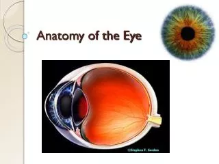
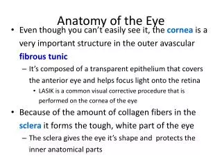
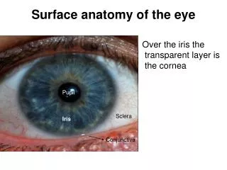
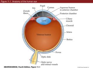
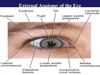
A couple key terms • Sclera: Outermost layer, gives eye it’s shape. • Cornea: The front layer of the sclera. Light hits this section first. • Extraocular muscles: The muscles that allow you to move your eye.
A couple key terms • Choroid: Second layer. Holds all the blood vessels responsible for getting blood to the eye. • Ciliary Body: Front of choroid. Muscles allow the lens to relax and adjust size. • Iris: That colored part of your eye! It’s all determined by pigment. Right in the middle is the pupil.
more anatomy • Dialator: This muscle in the iris is key for letting more light in the eye. Makes iris smaller and pupil larger • Sphincter: Another iris muscle that is the opposite of the dialator. Allows in less light, and makes the iris larger and pupil smaller.
Inner layer • The retina is the part of the eye that actually senses light • contains rods which take over in low light, and cones which take over color vision duties • In the back of the eye and center of the retina is the macula, which houses the fovea centralis, which allows us to see fine details.
View of the doctor • Vitreous humor fills a large section in the back of the eyeball • A smaller section houses aqueous humor, which is drained through the canal of Schlemm • Glaucoma results when that drainage is blocked up
More parts • The lens is a fascinating part that sharpens our vision • Ever had pink eye? That’s really called conjunctivitis or inflammation of the conjunctiva, a membrane that keeps our eyes moist.
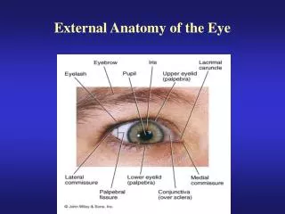
Protection • Ever wonder how our eyes remain relatively safe? • The bony structure called the orbital cavity provides protection; so does a cushion of fat • Our eyelids protect our eyes when we blink, and it spreads tears over the eyes (important for moisture) • Eyelashes and eyebrows keep dust and foreign objects out of the eye
We hope you learned something! • Special Thanks to HowStuffWorks.com