Download
1 / 100
1.01k likes | 1.16k Vues
Complex Carbohydrates Glycosaminoglycans(GAGs). Glycoproteins and proteoglycans. LECTURE OUTLINE. Differences between glycoproteins and proteoglycans Structures of glycoproteins and proteoglycans Functions of glycoproteins and proteoglycans

E N D
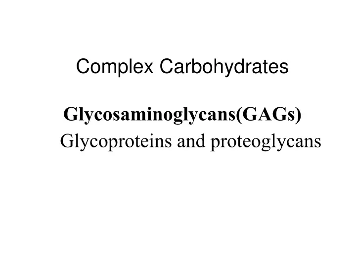
Complex CarbohydratesGlycosaminoglycans(GAGs) Glycoproteins and proteoglycans
LECTURE OUTLINE • Differences between glycoproteins and proteoglycans • Structures of glycoproteins and proteoglycans • Functions of glycoproteins and proteoglycans • Synthesis and degradation of glycoproteins and proteoglycans • Pathology related to glycoproteins and proteoglycans
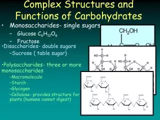
Differences Between Glycoproteins and Proteoglycans Proteins conjugated to saccharides lacking a serial repeat unit Glycoproteins Protein>>carbohydrate Carbohydrate>>protein Proteoglycans Proteins conjugated to polysaccharides with serial repeat units Glycosaminoglycans Mucopolysaccharides
Glycoprotein Glycoproteins are proteins that contain oligosaccharide chains (glycans) covalently attached to their polypeptide side-chains. The process of attaching the glycans is known as glycosalation. The sugar groups attached to glycoprotein can assist in protein folding or improve a proteins’ stability.
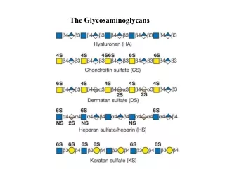
The disaccharide units contain either of two modified sugars, called amino sugarsN-acetylgalactosamine (GalNAc) or N-acetylglucosamine (GlcNAc), and an acidic sugar uronic acid such as glucuronic acid or iduronic acid. The amino group is usually acetylated.
In some glycosaminoglycans, one or more of the hydroxyls of the amino sugar is esterified with sulfate. The combination of these sulfate groups and the carboxylate groups of the uronic acid residues gives the glycosaminoglycans a very high density of negative charge. Keratan sulfate is an exception in which galactose is present, instead of an acidic sugar. Hyaluronic acid does not contain sulfate.
Structure of Glycosaminoglycans GAGs in the body are linked to core proteins ( except hyaluronic acid), forming proteoglycans (also called mucopolysaccharides). The GAGs extend perpendicularly from the core in a brush-like structure. e.g. in cartilage proteoglycan the GAGs present are chondriotin sulfate and keratan sulfate
Proteoglycan Aggregates Proteoglycan monomers associate with a molecule of hylauronic acid to form proteoglycan aggregates in association with linker proteins in a “bottle brush” arrangement.Association is not covalent but ionic between hyaluronic acid and the core protein. Stabilized by link proteins
Proteoglycan aggregate of the extracellular matrix One very long molecule of hyaluronan is associated noncovalently with about 100 molecules of the core protein aggrecan
Interactions between cells and the extracellular matrix with binding sites for both integrin and the proteoglycan
Linkage The linkage of GAGs to the protein core involves a specific trisaccharide composed of two galactose residues and a xylose residue (Gal-Gal-Xyl-O-CH2-protein).
The trisaccharide linker is coupled to the protein core through an O-glycosidic bond to a Serine residue in the protein. Some forms of keratan sulfates are linked to the protein core through an N-glycosidic bond. The protein cores of proteoglycans are rich in Serine and Threonine residues, which allows multiple GAG attachments.
STRUCTURE OF GLYCOPROTEINS One or more carbohydrate chains--covalently linked to a protein. The chains may be neutral or negatively charged. They are frequently branched. There are two types of glycosidic links: 1. O-glycosidic link O-glycosidic link between galactose or glucose and the hydroxyl group of hydroxylysine (i.e. collagen). Other O-linked glycoproteins have a glycosidic link between N-acetyl galactosamine and either serine or threonine (i.e. blood group substances and salivary mucins). 2. N-glycosidic link N-glycosidic links exist between N-acetylglucosamine and asparagine. There are two types: A. High mannose B. Complex. For example, in addition to mannose they may contain N-acetylglucosamine, galactose, fucose and N-acetylneuraminic acid (sialic acid)
Their core pentasaccharide is the same. In the complex form additional sugar residues are present: N-acetylglucosamine (GlcNAc) and N-acetylneuraminic acid (NANA). Fucose
Glycoproteins Glycoproteins are proteins that contain oligosaccharide (glycan) chains covalently attached to their polypeptide backbones. Glycoproteins occur in most organisms, from bacteria to humans. Their carbohydrate content ranges from 1% to over 85% by weight.
They differ from proteoglycans: Length of the chain is relatively short (usually 2-10 sugar residues) very long in GAGs. Do not have repeating disaccharide units. They are branched. May or may not be negatively charged.
The distinction between proteoglycans and glycoproteins resides in the level and types of carbohydrate modification.
Proteoglycans also contain the sugar glucuronic acid (GlcA). The carbohydrate modifications found in glycoproteins are rarely as complex as that of proteoglycans. Most proteins that are secreted, or bound to the plasma membrane, are modified by carbohydrate attachment. The part that is modified, in plasma membrane-bound proteins, is the extracellular portion of the protein.
Intracellular proteins are less frequently modified by carbohydrate attachment. However, the attachment of carbohydrate to intracellular proteins confers unique functional activities on these proteins
Structure of Glycoprotein The oligosaccharide components of glycoproteins is branched heteropolymers composed of D-hexoses, with the addition in some cases of neuraminic acid, and of L-fucose (6-deoxyhexose)
Some Functions of Glycoproteins _________________________________________________ Function Glycoprotein _________________________________________________ 1. Structural molecule Collagens/Arthritis 2. Lubricant Mucins/ Peptic ulcer 3. Transport molecule e.g. Transferrin, Ceruloplasmin 4. Immune system Immunoglobulins, Histocompatibility antigens, Blood group determinants 5. Hormone e.g. HCG, TSH 6. Enzymes e.g. Alkaline phosphatase 7. Blood clotting e.g. Fibrinogen 8. Blood groups 9. Cell surface recognition Lectins
Glycoproteins • Glycoproteins contain carbohydrate units covalently bonded to a polypeptide chain • antibodies are glycoproteins • carbohydrates play a role as antigenic determinants, the portions of the antigenic molecule that antibodies recognize and to which they bond.
Almost all the plasma proteins of humans—except albumin—are glycoproteins. For example, O-linked oligosaccharides on the surface of RBCs help provide the ABO blood group determinants Many proteins of cellular membranes contain substantial amounts of carbohydrate.
Blood Group Substances • Membranes of animal plasma cells have large numbers of relatively small carbohydrates bound to them: • these membrane-bound carbohydrates act as antigenic determinants • among the first antigenic determinants discovered were the blood group substances • in the ABO system, individuals are classified according to four blood types: A, B, AB, and O • at the cellular level, the biochemical basis for this classification is a group of relatively small membrane-bound carbohydrates
ABO Blood Classification • in type A, the nonreducing end is NAGal • in type B it is Gal • in type AB, both types are present • in Type O, neither of these terminal residues is present
lectin-ligand interactions in lymphocyte movement to the site of an infection Stronger interaction near the site of inflammation
Helicobacter pylori Interaction between a bacterial surface lectin and an oligosaccharide of the gastric epithelium
The extracellular space in animal tissues is filled with a gel-like material, the extracellular matrix, also called ground substance, which holds the cells of a tissue together and provides a porous pathway for the diffusion of nutrients and oxygen to individual cells.
Epithelial cells extra-cellularmatrix Underlying cells
The extracellular matrix is composed of an interlocking meshwork of heteropolysaccharides and fibrous proteins. Heteropolysaccharides in the body are the glycosaminoglycans (GAGs). These molecules are long unbranched polysaccharides containing a repeating disaccharide unit.
GAGs are highly negatively charged molecules, with extended conformation that imparts high viscosity to the solution. GAGs are located primarily on the surface of cells or in the extracellular matrix (ECM).
Along with the high viscosity of GAGs comes low compressibility, which makes these molecules ideal for a lubricating fluid in the joints. At the same time, their rigidity provides structural integrity to cells and provides passageways between cells, allowing for cell migration.
Hyaluronic acid • Hyaluronic acid is unique among the GAGs in that it does not contain any sulfate and is not found covalently attached to proteins as a proteoglycan. • It is, however, a component of non-covalently formed complexes with proteoglycans in the ECM.
Only GAG present both in animals and bacteria. Found in synovial fluid, vitreous humor, ECM of loose connective tissue Umbilical cord Cartilage
b-1,3 b-1,4 Glycosaminoglycans GlcUA GlcNAc No protein link No sulfate b-1,3 glycosidic linkage Hyaluronate
Specific function: 1.Hyaluronic acid is especially high in concentration in embryonic tissues and is thought to play an important role in permitting cell migration during morphogenesis and wound repair. 2. Act as lubricators and shock absorbers.
Association with major diseases: Hyaluronic acid may be important in permitting tumor cells to migrate through the ECM. Tumor cells can induce fibroblasts to synthesize greatly increased amounts of this GAG, thereby perhaps facilitating their own spread.
b-1,3 b-1,4 Glycosaminoglycans GlcUA GalNAc GlcUA-Gal-Gal-Xyl-O-Ser link Sulfate at 4 or 6 C of GalNAc b-1,3 glycosidic linkage Chondroitin sulfate
Chondroitin sulfate Most abundant GAG Cartilage (bind collagen and hold the fibers strongly) Tendons Ligaments Heart valves
a-1,4 a-1,4 Glycosaminoglycans GlcUA GlcNAc GlcN and GlcUA or IdUA N and O sulfate (C2,3,6) a-1,4 glycosidic linkage Heparin > NAc < N and O sulfate Heparan sulfate