Download
1 / 34
360 likes | 847 Vues
FACIAL NERVE PRESERVATION IN VESTIBULAR SCHWANNOMAS. Contents . Introduction Anatomy of facial nerve Imaging Intra-op landmarks and intra-op nerve monitoring Grading of facial palsy Factors associated with preservation of facial nerve Microsurgical resection Radiosurgery

E N D
Contents • Introduction • Anatomy of facial nerve • Imaging • Intra-op landmarks and intra-op nerve monitoring • Grading of facial palsy • Factors associated with preservation of facial nerve • Microsurgical resection • Radiosurgery • Facial nerve re-animation techniques • Conclusions
Introduction • Vestibular schwannomas- Most common of intracranial schwannomas. • Arise from the transition zone of myelin at the porusacousticus (Obersteiner- Reidlich zone) • MC arise from the inferior vestibular nerve • Peak incidence in 4th -6th decade • Sporadic/Familial
Grading • Koos: (Grade 1-4) upto 1, 2, 3 and >3 cm (intracanalicular+ cisternal) • Ojemann: (small, med, large)<2, 2-3 >3cm (intracisternal) • Samii: >3×2cm large, rest small. (both intra + extrameatal), also T1, T2, T3ab, T4ab • Shekhar: (small, med, large) <2, 2-3.9, >3.9 cm (only intracisternal)
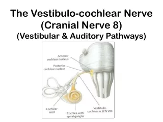
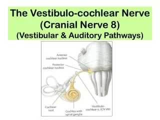
Facial nerve • Seventh cranial nerve • Motor and sensory components (motor- 70%, sensory-30%) • Sensory part also called nerve of Wrisberg • Branchiomotor- supplies muscles of second branchial arch
Structures supplied • Motor • Muscles of facial expression • Muscles of scalp and ear • Buccinator, stapedius, stylohyoid, posterior belly of digastric, platysma • Parasympathetic secretory fibers to sublingual and submandibular salivary glands, lacrimal gland and mucous membranes of oral and nasal cavities
Sensory • Taste- anterior 2/3 rd of tongue • Exteroceptive- eardrum and EAC • Proprioceptive- muscles it supplies • General visceral sensation- salivary glands and mucosa of nose and pharynx • Anatomically, motor part is separate from the sensory and parasympathetic
3 parts • Intracranial part- Pons to IAC ( 15-17 mm) • Intratemporal part- IAC to stylomastoid foramen • Meatal segment (8-10 mm)- within meatus • Labyrinthine segment- from fundus of meatus to geniculate ganglion; here, facial nerve has the narrowest diameter(0.61-0.68mm) and shortest segment (4 mm) • Tympanic/ horizontal segment- from geniculate ganglion to just above the pyramidal eminence ( 11mm) • Mastoid/ vertical segment- from pyramid to stylomastoid foramen • Extra-cranial part- from stylomastoid foramen to termination of branches
Nervusintermedius(Nerve of Wrisberg) • Sensory and parasympathetic division • Preganglionic parasympathetic fibres to • Submaxillary ganglion ( to sublingual and submandibular glands) • Pterigopalatine ganglion ( to lacrimal, palatal and nasal glands) • Also receives sensory fibres from geniculate ganglion
Facial nerve identification-Imaging • Routine T2WI not sufficient for identifying facial nerve • DTI based tractography can be utilized to know the relation of facial nerve (also other cranial nerves) to the tumour • Gerganov et al, Diffusion tensor imaging–based fiber tracking for prediction of the position of the facial nerve in relation to large vestibular schwannomas. JNS Dec 2011- 22 patients- DTI to surgical correlation was 90% • Chen et al -DTI with tractography- 3 patients; could identify facial and trigeminal nerves. Neurosurgery Apr 2011-3 patients-only imaging identification.
History • Sir Charles Ballance first successfully resected an acoustic neuroma in 1894 • Harvey Cushing- advocated subtotal removal • Walter Dandy (1925)- first surgeon to totally resect acoustic tumours successfully • Dandy himself wrote that "paralysis of the facial nerve must usually be accepted as a necessary sequel of the operation.“ • Cairns (1931)- first surgeon to document facial nerve function preservation • Olivecrona (1940)- Performed surgeries by observing facial twitches to guide tumour resection
Goal of surgery has changed from prolongation of life to cranial nerve function preservation. Loss of facial nerve function is a debilitating and psychologically devastating condition. According to the Acoustic Neuroma Association, facial nerve dysfunction remains the number one concern among patients undergoing cerebellopontine angle surgery.
Facial nerve palsy-Pathogenesis • Most common cause of postoperative facial nerve palsy is direct trauma or nerve stretching during surgery (mostly neuropraxia/ axonotmesis) • Devascularization of nerve segments that are effaced by large tumors. • Thermal injury (both hot and cold)
How to minimize? • Initial debulking f/b dissection • Dissect the tumour from the nerve and not vice-versa • Excessive pressure on facial nerve to be avoided • Cottoinoids and microsuction devices to be used • Sharp dissection is a must until clear plane is identified • Avoid excessive cerebellar retraction to avoid undue tension on the nerve
Arterial supply of facial nerve • Labyrinthine artery branch of AICA • Greater superficial petrosal branch of MMA • Stylomastoid branch of ECA • Maintaining blood supply is critical • Avoid inadvertent vascular injury • Blunt dissection near all vascular structures • Maintain arachnoid plane • Topical papaverine after resection to prevent vasospasm
Thermal injury • Both hot and cold can lead to facial n paresis • Lasers (CO2) can cause permanent damage • Caution while using bipolar cautery near nerves • During drilling of IAM, continuous warm saline irrigation is recommended. • Overly cold irrigation may "stun" the nerve and is avoidable with use of warmed saline solutions.
If facial nerve disrupted during surgery • Immediate repair is advisable • Direct proximal to distal anastomosis • Intracranial- intra-temporal (by drilling the temporal bone) • Intracranial- extracranial techniques • If no function returns- then facial reanimation • Not later than 1 year
Intra-op facial nerve monitoring • Olivecrona was the first to monitor facial function during surgery-1950 • Practical neurophysiologic monitoring first introduced by Delgado in 1979 • Now considered a standard in VS surgery. • VII nv monitoring • EMG monitoring of muscles innervated by VII nv • Displayed on an oscilloscope connected to an audio amplifier • Statistically significant difference in anatomical & functional VII nv preservation • Enables the surgeon to obtain instantaneous feedback on facial nerve firing during tumor dissection • Stimulation of the facial nerve at the brainstem with a threshold <0.05 mA predicts good facial nerve outcome.
Allows definitive and early identification of facial nerve and thereby speeds up the dissection • Reduced the operative times substantially, although not enhanced the facial nerve preservation substantially • Sampath et al: Facial nerve injury in acoustic neuroma (vestibular schwannoma) surgery: etiology and prevention. Neurosurg Focus 1998 • Stimulation should be used liberally throughout the operation. • Electrical status of the nerve to be always determined immediately before closure by stimulation at the brainstem and the entire course.
Immediately postoperatively, 75% of the 0.1 mA threshold group, 42% of the 0.2 mA group and 18% of the >= 0.3 mA group had good (Grade I or II) facial nerve function. • One year postoperatively, 90% of the 0.1 mA group, 58% of the 0.2 mA group and 41% of the >= 0.3 mA group had Grade I or II function. • Statistically significant breakpoint of 0.2 mA was found to predict good postoperative facial function
AIIMS data • Facial nerve anatomically preserved in 78%, last follow up- 82% patients showed acceptable facial function. • GTR in 24.2%, NTR 47.2% and STR 28.6%. • Microsurgical management of giant acoustic neuromas: An institutional series of 400 cases: Sinha S, Sharma B S, Asian Journal of Neurosurgery 2008
Literature review • Microsurgical resection: (78-85%) • Age < 65 yrs (84% v/s 71%) • Approach: Middle fossa approach (85%)> Translabyrinthine (81%)> Suboccipital(78%) • Tumour size: < 20 mm (90% v/s 67%) • Use of intra-op nerve monitoring (76% v/s 71%) • Sughrue ME et al: Preservation of facial nerve function after resection of vestibular schwannoma. Br J Neurosurg 2010 Dec • 79 studies, 11873 pts • Grade 3 or higher facial palsy were excluded.
Radiosurgery: (96.2%) • Tumour volume- <1.5 cm3 • Marginal radiation dose</=13 Gy • Age< 60 yrs • Yang I et al: Facial nerve preservation after vestibular schwannoma Gamma Knife radiosurgery. J Neurooncol 2009 May • 23 studies, 2200 pts. • Average F/U-54.1+/- 31.3 mts
Facial nerve sparing approach for VS • Small tumours (<2.2 cm3)- Primary GKRS • Larger tumours (>3 cm)/ severe symptoms- Primary microsurgical resection • GTR- if feasible and facial nerve not at risk (by IOP monitoring) • Or else STR f/b GKRS for significant residual/recurrent tumour. • Rate of preservation-around 97% • Haque R et al: Efficacy of facial nerve–sparing approach in patients with vestibular schwannomas. JNS Nov 2011.
Facial nerve re-animation • Refer to interventions that restore facial symmetry, resting tone, voluntary movement, or a combination of these. • Several broad categories of facial reanimation techniques exist • Reinnervation techniques • Muscle transfers and • Static procedures
Dynamic procedures- improve facial tone & motor function Primary nerve repair Nerve grafting Neuromuscular pedicle grafts Regional muscle Transposition Microvascular muscle transfers Static procedures- - add support and symmetry to the patient’s face at rest - supplement results of nerve grafting/ dynamic procedures Gold weight implantation in upper eyelid Palpebral sling placement Lower lid ectropion correction
Re-innervation techniques • Also termed nerve substitution techniques • Provide neural input to the distal facial nerve through motor nerves other than the ipsilateral facial nerve • Nerves used: • Hypoglossal nerve-MC used • C/L facial nerve • Others • Spinal accessory • Trigeminal nerve • Glossopharyngeal nerve
Muscle transposition techniques • Indicated in cases of significant atrophy of facial musculature • Muscles used • Temporalis- MC used • Others • Masseter • Digastric • Free muscle transfers • Gracilis
Static facial reanimation procedures • Indications: • Patients who are poor candidates for prolonged general anesthesia for medical reasons • Patients with a poor prognosis in whom reanimation over a long time is not appropriate • Dynamic reanimation failures. • Patients with partial recovery following Bell’s palsy, Ramsay Hunt syndrome, or other conditions leading to aberrant regeneration
Nasal valve repair-for dilator nares paralysis • Static procedures for paralyzed eyelids • Lateral tarsorrhaphy ( ? cosmetic concern) • Gold weight implantation in upper eyelid – to restore eyelid closure • Palpebral sling placement • Procedure to correct lower lid ectropion – implant a piece of auricular cartilage in the lower eyelid
Conclusions • Goals of surgery- changed from prolongation of life to preservation of cranial nerve function • Sound anatomical knowledge, good microsurgical techniques, especially maintenance of anatomical planes- very crucial • Pre-op tumour size- significant factor in facial nerve outcome • Use of intra-op nerve monitoring- valuable adjunct in acoustic tumour surgeries. • Facial palsy complications to be dealt with aggressively including reanimation techniques