Download
1 / 24
240 likes | 422 Vues
Chapter two Arthrology. Department of Anatomy 张正洪 博士 / 副教授. Classification. There are two major types of articulations ( or joints ) . Continuous joints/immovable joints Fibrous joints : bones are united by fibrous connective tissue

E N D
Chapter two Arthrology Department of Anatomy 张正洪 博士/副教授
Classification There are two major types of articulations (or joints). • Continuous joints/immovable joints • Fibrous joints : bones are united by fibrous connective tissue • Cartilaginous joints : bones are united by cartilage • Synosteosis • Discontinuous joints -synovial joints
Synovial joints Basic structures • Articular surface: covered byarticular cartilage • Articular capsule • Fibrous membrane • Synovial membrane • Articular cavity: containing a trace of synovial fluid; subatmospheric pressure in it
Accessory structures • Ligaments(lig.): extra- and intracapsular ligaments • Articular disc and articular labrum • Synovial fold andsynovial bursa
Movements of joints • Translation • Flexion and extension • Adduction and abduction • Rotation • Medial and lateral rotation • Pronation and supination • Circumduction
Classification of synovial joints • Uniaxial joints • Biaxial joints • Multiaxial joints
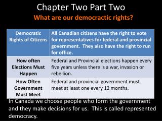
Articulations of Bones of Trunk The vertebral column consists of 24 vertebrae, the sacrum, and the coccyx.
Joints of the vertebral bodies Intervertebral discs between bodies of adjacent vertebrae, composedof: • Nucleus pulposus, an inner soft, pulpy, highly elastic structure (gelatinous core ) • Annulus fibrosus an outer fibrous ring consisting of fibrocartilage
Anterior longitudinal ligament • Maintains stability of the intervertebral disc and prevents hyperextension of the vertebral column Posterior longitudinal ligament • Prevents hyperflexion of the vertebral column and posterior protrusion of the discs
Joints of the vertebral arches • Ligamenta flava ― elastic ligament, unite laminae of adjacent vertebrae, and complete the posterior wall of vertebral canal; tend to prevent hyperflexion of the vertebral column
Interspinal ligament • Supraspinal ligament • Ligamentum nuchae • Intertansverse ligament • Zygapophysial joint
Atlantooccipital joint • Between superior articulating surfaces of atlas and occipital condyles • Supported by membrances and ligaments that join occipital bone and atlas • Action ― nodding of head, lateral tilting of head
Atlantoaxial joint • Three synovial joints between atlas and axis • Supported by ligaments • Action ― allow atlas (and head) to pivot on the axis and vertebral column
View of bertebral column as a whole Anterior view Posterior view Lateral view: • Cervical curvature • Thoracic curvature • Lumbar curvature • Sacral curvature
Movement of the vertebral column • flexion • extension • lateral flexion • Rotation • circumduction
Thoracic cage (thorax) Composition Bones― consists of twelve thoracic vertebraes, twelve pairs of ribs and costal cartilages, and sternum
Joints • Costovertebral joints • Joints of costal head • Costotransverse joints
Sternocostal joints • Sternocostal synchondrosis of first rib • Sternocostal joints • Interchondral joints: between costal cartilages 8, 9, and 10 to form the costal arch
General features of thoracic cage • Roughly cone-shape, narrow above and broad below, flattened from before-backwards, longer behind than in front • Inlet of thorax • Outlet of thorax • Infrasternal angle • Intercostal spaces
Function: • protects the organs in the thoracic cavity and upper abdominal cavity; • plays a vital role in the process of breathing Inspiration Expiration
Joints of skull • Continuous joints: sutures, synchondrosis or synosteosis
Temporomandibular joint • Articular surfaces • Capsule: thin and lax in front and behind; strengthened by the lateral ligament • Articular disc • Movement