Download
1 / 38
380 likes | 510 Vues
Objective. To present a case of a Hemophagocytic Lymphohistiocytosis (HLH). General Data. BM 6 year old Male from Bacoor, Cavite. Chief complaint. FEVER. History of Present Illness. (+) fever (Tmax: 40degrees C) (+) occasional cough (+) headache and joint pains

E N D
Objective • To present a case of a Hemophagocytic Lymphohistiocytosis (HLH)
General Data • BM • 6 year old • Male • from Bacoor, Cavite
Chief complaint FEVER
History of Present Illness (+) fever (Tmax: 40degrees C) (+) occasional cough (+) headache and joint pains Paracetamol for the fever 5 days PTA (+) persistence of fever Consult done at St. Dominic Hospital CBC: Hgb 122/ Hct 37/ WBC 7.27/ neutro 63/ lymph 24/ platelet 289 UA: normal Dengue NS1 negative Impression: Upper respiratory tract infection Clarithromycin 3 days PTA
History of Present Illness (+) persistence of fever (+) abdominal pain, nausea, myalgia Follow-up consult St. Dominic hospital rpt CBC: Hgb 110/ Hct 35/ wbc 1.7/ neutro 36/ lymph 58/ platelet 95 Typhidot: negative Advised admission, but opted transfer to our institution On the day of admission ADMISSION
Past Medical History • no previous hospitalization • (+) asthma • no known allergies
Family History • (-) asthma, allergies • (-) cancer • (-) DM, HPN
Immunization History BCG 1 dose Hepatitis B 3 doses DPT 3 doses OPV 3 doses Pneumococcal none Rotavirus none Hepatitis A 1 dose Typhoid none
Physical exam • Awake, weak-looking , not in cardiorespiratory distress • BP: 90/60mmHg CR: 98bpm RR: 24 cpm T: 39.6 C • Weight: 19.8 kg Height 131 cm • pink palpebral conjunctivae, anicteric sclerae • moist oral mucosa, no tonsillopharyngeal congestion • good air entry, clear breath sounds • regular cardiac rhythm, grade 2/6 systolic murmur on left parasternal border • soft abdomen, nontender • full and equal pulses, CRT <2sec
CBC done: • Dengue blot negative • Admitted as a case of Systemic Viral Illness r/o Dengue fever • IV hydration, Paracetamol
Course in the Wards • Problems: • (+) persistence of fever • occasional cough • increased effort in breathing • with episodes of abdominal pain • cardiac findings: Grade 2/6 systolic murmur on left parasternal border • respiratory: RR 30-60s, harsh breath sounds, no rales, no wheezes • abdominal findings: soft abdomen, liver edge palpable 3-4 cm below the subcostal margin
Course in the Wards • Infectious
Course in the Wards • Cardiac: ECG: Normal sinus rhythm, normal axis
Course in the Wards • Respiratory: CHEST XRAY Bilateral Interstitial Pneumonitis) Ceftriaxone 1.5g/IV OD
Course in the Wards • Abdomen: PLAIN ABDOMINAL XRAY: No localizing signs in the abdominal tissues • ABDOMINAL ULTRASOUND prominent sized liver, mild to moderate ascites and pleural effusion
Course in the Wards Sodium correction Albumin transfusion
Course in the Wards • REFERRALS • IDS: Cannot totally rule out Dengue fever; HLH • GI : Systemic Viral Illness, Postinfectious hepatitis • HEMATOLOGY: t/c HLH
Transfer to PICU Meropenem 750 mg/IV every 8h (113.6mkd) • prolonged fever • tachypnea RR 50s and labored breathing • tachycardia HR 110-120s bpm • still with episodes of abdominal pain O2 support 2lpm NC 2d-echo: pericardial effusion, mild MR, mild TR Carditis with diastolic dysfunction t/c SEPSIS rule out HLH
Transfer to PICU pRBC transfusion • anemia, leukopenia, thrombocytopenia • electrolyte imbalance (hypokalemia, hypocalcemia) • fluid spacing (pleural effusion, ascites) • deranged liver function tests K correction Calcium gluconate Furosemide
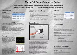
Diagnostic Criteria for HLH Text Treatment Protocol of the 2nd International HLH Study, 2004.)From Verbsky JW, Grossman WJ: Hemophagocytic lymphohistiocytosis: diagnosis, pathophysiology, treatment, and future perspectives, Ann Med 38:20–31, 2006, p 21, Table 1 Retrieved from Nelsons Pediatrics 19th ed.
At the PICU • CT Abdomen with Contrast: • Prominent sized liver and spleen • Liver 13.5 cm right midclavicular line • Spleen 8.2 x 8.8 x 4.3 cm • Ascites • Bilateral pleural effusion, right more than left
At the PICU • Triglycerides 460.20 mg/dl (nv 0-150) • Fibrinogen 136.2 mg/dl (nv 180-350) • Ferritin >40,000 ng/ml (nv 8.80 - 184.7)
At the PICU • BMA: Hemagophagocyte - 1 • for flow cytometry
Hemophagocytic Lymphohistiocytosis • also called “hemophagocytic syndrome (HPS) • a nonmalignant proliferative disorder that affects the antigen-processing macrophages and that results in uncontrolled hemophagocytosis and upregulation of inflammatory cytokines impaired natural killer (NK) cell function and other defects Palazzi D L et al. Hemophagocytic Syndrome in Children: An Important Diagnostic Consideration in Fever of Unknown Origin. Clin Infect Dis. 2003;36:306-312
Hemophagocytic Lymphohistiocytosis • A potentially fatal disorder of children and adults due to cytokine dysfunction, resulting in uncontrolled accumulation of activated T-lymphocytes and activated histiocytes (macrophages) in many organs. • HLH may be familial, associated with a number of different infections, autoimmune disorders, or coincident with a number of malignancies. HLH-2004: Diagnostic and therapeutic guidelines for hemophagocytic lymphohistiocytosis.AU Henter JI, Horne A, AricóM, Egeler RM, Filipovich AH, Imashuku S, Ladisch S, McClain K, Webb D, Winiarski J, Janka G SO. Pediatr Blood Cancer. 2007;48(2):124.
Primary HLH • Familial hemophagocytic lymphohistiocytosis (FHLH) (familial or sporadic): • an autosomal recessive disease that affects immune regulation • Nonfamilial HLH: • develop from marked immunological activation during viral, bacterial, and parasitic infections • may also be associated with malignancies, prolonged administration of lipids, rheumatoid arthritis (macrophage activation syndrome), immune deficiencies associated with cytotoxic T- and/or Nkcell dysfunction such as DiGeorge syndrome (del 22q11.2), Chédiak–Higashi syndrome, Griscelli syndrome,* X-linked lymphoproliferative disease (XLP), and lysinuric protein intolerance (LPI). Manual of Pediatric Hematology and Oncology 4th ed . P. Lanzkowsky (Elsevier, 2005).
Secondary HLH • A reactive disorder causing strong immunologic activation often resulting from severe bacterial or parasitic infection • Infection-associated HPS IAHS] • Viral infection (VAHS) • Malignancy (MAHS) • Use of drugs (phenytoin) • Prolonged administration of parenteral nutrition involving soluble lipids Palazzi D L et al. Hemophagocytic Syndrome in Children: An Important Diagnostic Consideration in Fever of Unknown Origin. Clin Infect Dis. 2003;36:306-312
Infection-Associated Hemophagocytic Lymphohistiocytosis • NK-cell activity in IAHLH patients is reconstituted as soon as the infection is cleared • decreased or absent NK cells are found more often in FHLH • Viruses: • Epstein–Barr virus, human herpes virus 6 (HHV-6), cytomegalovirus (CMV) (most common of the viruses), adenovirus, parvovirus, varicella zoster, herpes simplex virus (HSV), Q-fever virus, and measles • Treatment • EBV–related IAHLH: etoposide and immunoglobulin treatment • Other infections: antibiotics for bacterial infections, antiviral drugs for viruses, in addition to corticosteroids and/or etoposide. • Patients with persistent HLH may require FHLH treatment and HSCT. • Patients with resolved disease may discontinue therapy at 8 weeks. If the syndrome recurs therapy should be restarted and HSCT should be employed.
Clinical features • Age of onset: Less than 1 year of age (70% of cases) *no known upper age limit for the onset of disease • Signs and symptoms of FHLH Most common early findings: a. Fever (91%) b. Splenomegaly (98%) c. Hepatomegaly (94%) Manual of Pediatric Hematology and Oncology 4th ed . P. Lanzkowsky (Elsevier, 2005).
The most common reason for hospital admission was fever with or without additional symptoms or signs. Hemophagocytic Syndrome in Children: An Important Diagnostic Consideration in Fever of Unknown OriginDebra L. Palazzi,1 Kenneth L. McClain,2 and Sheldon L. Kaplan11Infectious Diseases Section and 2Section of Hematology and Oncology, Department of Pediatrics, Baylor College of Medicineand Texas Children’s Hospital, Houston, Texas (2003)
Clinical features Other findings: a. Lymph node enlargement (17%) b. Skin rash (6%) c. Neurologic abnormalities (20%) • Neurologic: irritability, bulging fontanel, neck stiffness, hypotonia, hypertonia, convulsions, cranial nerve palsies, ataxia, hemiplegia, blindness, and unconsciousness d. Multisystem involvement: lungs, bone marrow, and leptomeninges. • Occasionally: ocular, heart, skeletal muscles, and kidney Manual of Pediatric Hematology and Oncology 4th ed . P. Lanzkowsky (Elsevier, 2005).
Diagnostic Criteria for HLH Text Treatment Protocol of the 2nd International HLH Study, 2004.)From Verbsky JW, Grossman WJ: Hemophagocytic lymphohistiocytosis: diagnosis, pathophysiology, treatment, and future perspectives, Ann Med 38:20–31, 2006, p 21, Table 1 Retrieved from Nelsons Pediatrics 19th ed.
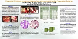
Pathologic findings —result from the aggressive proliferation of normal histiocytes and T-lymphocytes in various tissues. Hemophagocytosis of red cells (erythrophagocytosis), other white blood cells, or platelets in the bone marrow, spleen, or lymph nodes is the key diagnostic finding. Bone marrow from a child with hemophagocytic syndrome, secondary to Epstein-Barr virus infection. Reactive histiocytes show phagocytosis of nucleated red blood cells (red arrows) and platelets (black arrows). Wright-Giemsa stain
Treatment • Dexamethasone 10 mg/m2/day for 2 weeks followed by a decrease every 2 weeks to 5 mg/m2, 2.5 mg/m2, and 1.25 mg/m2 for a total of 6 weeks • Etoposide IV (150 mg/m2 IV 2-hour infusions daily) twice weekly for 2 weeks, then weekly • Cyclosporine A 3–5 mg/kg/day by continuous IV infusion starting week 8 to reach a blood trough level of 150–200 ng/mL and switching to oral administration of 6–10 mg/kg/day in two divided doses • Intrathecal methotrexate (IT MTX), age-adjusted doses of IT MTX weekly for 3–6 weeks as follows if there are progressive neurologic symptoms or if abnormalcells persist in the CSF • Allogeneic stem cell transplantation (BMT) after cytotoxic chemotherapy for all patients with familial disease or those with persistent nonfamilial disease. Manual of Pediatric Hematology and Oncology 4th ed . P. Lanzkowsky (Elsevier, 2005). Modern management of children with haemophagocytic lymphohistiocytosis. AU Janka GE, Schneider EM SO Br. J Haematol. 2004;124(1):4.
Treatment • The standard of care in 2011 for HLH patients being treated outside of a therapeutic research trial is treatment with dexamethasoneand etoposide, as outlined in the HLH-94 trial. • Cyclosporine may be added (initial dose 6mg/kg per day, divided in two daily doses; target trough levels 200 mcg/L), but the benefit of its use during the initial eight-week induction period is not yet proven and its use has been associated with posterior reversible encephalopathy syndrome (PRES) • In the HLH-2004 research protocol, cyclosporine will be started on day one. Modern management of children with haemophagocytic lymphohistiocytosis. AU Janka GE, Schneider EM SO Br. J Haematol. 2004;124(1):4.
Prognosis • Without treatment, FHLH is usually rapidly fatal, with a median survival of about 2 months. • Chemotherapy and immunosuppressive therapy may prolong survival in FHLH but only stem cell transplantation may be curative. • Patients with known familial disease or severe or persistent acquired disease should receive hematopoietic stem cell transplantation (HSCT). The 3-year actuarial survival in familial HLH with this approach has been reported as 51% ± 20%.