Download
1 / 27
280 likes | 408 Vues
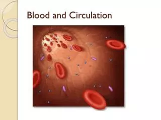
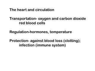
The heart and circulation. Structure of the heart Regulation of heart activity The circulatory system- cardiovascular and lymphatic. Structure of the heart Two atria, two ventricles Atria receive blood from venous system Ventricles pump blood into arterial system

E N D
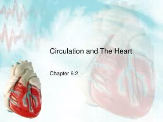
The heart and circulation Structure of the heart Regulation of heart activity The circulatory system- cardiovascular and lymphatic
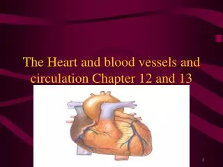
Structure of the heart Two atria, two ventricles Atria receive blood from venous system Ventricles pump blood into arterial system Septum separates right from left side
A double pump: pulmonary and systemic circulation
Valves embedded in fibrous skeleton AV valve between right atrium and ventricle- tricuspid valve AV valve between left atrium and ventricle bicuspid (mitral) valve Semilunar valves at base of pulmonary artery and aorta
Cardiac cycle and heart sounds Contraction- systole Relaxation- diastole Atria contract simultaneously Then ventricles contract- with a little overlap Stroke volume- amount of blood ejected from ventricles during systole end-systolic volume- what’s left
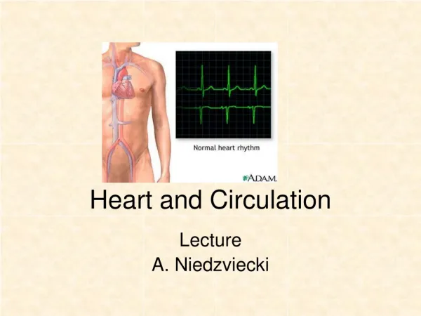
Electrical activity of the heart Myocardial cells beat automatically Action potential is usually originated in sinoatrial node Spontaneous depolarization (pacemaker potential) diffusion of calcium through slow channels threshold- fast calcium channels open, voltage regulated sodium channels open repolarization produced through diffusion of potassium
Other parts of the heart can produce pacemaker potentials Depolarize more slowly than SA node; usually stimulated by action potentials from SA node before they could start their own pacemaker potentials “ectopic pacemakers” can set a rhythm if SA node conduction is blocked; pace will be slower
Heart muscle cannot sustain contraction Long refractory periods- heart cannot be stimulated until it has relaxed from previous contraction Arrhythmias- something affects the cardiac cycle; treatment depends on what it is Fast Na channel blockers Slow Ca channel blockers -adrenergic receptor blockers
Blood vessels- arteries and veins Arteries, arterioles, capillaries Veins and venules Arteries are more muscular Veins have valves
Capillaries deliver blood to cells Specialized types of capillaries in different organs Fenestrated- kidneys, endocrine glands, intestines Discontinuous- bone marrow, liver and spleen Continuous- everywhere else
Veins Veins can expand to accommodate increasing amounts of blood; arteries can’t Venous pressure is low compared to arterial pressure Blood flow through veins is facilitated by: contraction of skeletal muscles valves that prevent backflow
Atherosclerosis • Plaques block blood vessels • Macrophages accumulate (“fatty streaks”) • Inflammatory mechanism that accumulates damage • Vasodilation function can be disrupted
LDL, HDL, and cholesterol • Cholesterol is carried to liver by LDL • Recycled via LDL receptors on liver cells • LDL (and cholesterol) can accumulate in blood • HDL carries cholesterol away from arterial walls • High HDL levels are beneficial
Arrhythmias Bradycardia- slow rate (less than 60 bpm) Tachycardia- fast rate (more than 100 bpm) Can occur normally; is abnormal if rate increases during rest (ectopic pacemakers) Flutters- extremely rapid contractions Fibrillation- different groups of fibers are activated so coordinated pumping of chambers is not possible
Lymphatic system Fluid transport from tissues Fat transport from intestines Immune response
Summary • “Double pump” enables heart to deliver oxygenated blood to the body- and recirculate it • Valves regulate blood movement through the heart • Electrical activity can be measured and monitored • Arterial system delivers blood to the body, and the venous system returns it to the heart • Lymphatic system helps regulate fluid levels among the body compartments