Download
1 / 29
320 likes | 1.09k Vues
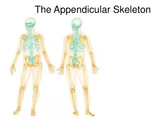
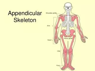
The Appendicular Skeleton Exercise 11 Page:145. Lab 5. Appendicular Skeleton. The appendicular skeleton is made up of the bones of the limbs and their girdles Pectoral girdles attach the upper limbs to the body trunk Pelvic girdle secures the lower limbs.

E N D
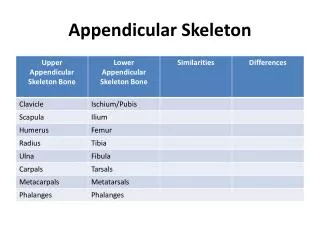
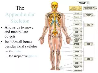
Appendicular Skeleton • The appendicular skeleton is made up of the bones of the limbs and their girdles • Pectoral girdles attach the upper limbs to the body trunk • Pelvic girdle secures the lower limbs
Pectoral Girdles (Shoulder Girdles) • The pectoral girdles consist of the anterior clavicles and the posterior scapulae • They attach the upper limbs to the axial skeleton in a manner that allows for maximum movement • They provide attachment points for muscles that move the upper limbs
Pectoral Girdles (Shoulder Girdles) Figure 7.22a
Clavicles (Collarbones) Figure 7.22b, c
Scapulae (Shoulder Blades) Figure 7.22d, e
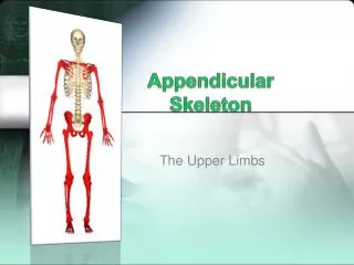
The Upper Limb • The upper limb consists of the arm (brachium), forearm (antebrachium),and hand (manus) • Thirty-seven bones form the skeletal framework of each upper limb
Arm Humerus • The humerus is the sole bone of the arm • It articulates with the scapula at the shoulder, and the radius and ulna at the elbow
Forearm Radius and Ulna The bones of the forearm are the radius and ulna. They articulate proximally with the humerus and distally with the wrist bones They also articulate with each other proximally and distally at small radioulnar joints. Interosseous membrane connects the two bones along their entire length Figure 7.24
Hand • Skeleton of the hand contains wrist bones ( 8 carpals), bones of the palm (metacarpals), and bones of the fingers (phalanges) Figure 7.26a
Pelvic Girdle (Hip) • The hip is formed by a pair of hip bones (os coxae, or coxal) • Together with the sacrum and the coccyx, these bones form the bony pelvis • The pelvis • Attaches the lower limbs to the axial skeleton with the strongest ligaments of the body • Transmits weight of the upper body to the lower limbs • Supports the visceral organs of the pelvis
Ilium • The ilium is a large flaring bone that forms the superior region of the coxal bone • It consists of a body and a superior winglike portion called the ala • The broad posterolateral surface is called the gluteal surface • The auricular surface articulates with the sacrum (sacroiliac joint) • Major markings include the iliac crests, four spines, greater sciatic notch, iliac fossa, arcuate line, and the pelvic brim
Ilium: Lateral View Figure 7.27b
Ilium: Medial View Figure 7.27c
Ischium • The ischium forms the posteroinferior part of the hip bone • The thick body articulates with the ilium, and the thinner ramus articulates with the pubis • Major markings include the ischial spine, lesser sciatic notch, and the ischial tuberosity
Pubis • The pubic bone forms the anterior portion of the hip bone • It articulates with the ischium and the ilium • Major markings include superior and inferior rami, the pubic crest, pubic tubercle, pubic arch, pubic symphysis, and obturator foramen (along with ilium and ischium)
Pubis: Lateral View Figure 7.27b
Comparison of Male and Female Pelvic Structure • Female pelvis • Tilted forward, adapted for childbearing • True pelvis defines birth canal • Cavity of the true pelvis is broad, shallow, and has greater capacity • Male pelvis • -Tilted less forward • -Adapted for support of heavier male build and stronger muscles • -Cavity of true pelvis is narrow and deep
Comparison of Male and Female Pelvic Structure Image from Table 7.4
The Lower Limb • The three segments of the lower limb are the thigh, leg, and foot • They carry the weight of the erect body, and are subjected to exceptional forces when one jumps or runs
Femur • The sole bone of the thigh is the femur, the largest and strongest bone in the body • It articulates proximally with the hip and distally with the tibia and fibula
Leg Tibia and Fibula • The tibia and fibula form the skeleton of the leg • They are connected to each other by the interosseous membrane • They articulate with the femur proximally and with the ankle bones distally • They also articulate with each other via the immovable tibiofibular joints
Foot • The skeleton of the foot includes the tarsus, metatarsus, and the phalanges (toes) • The foot supports body weight and acts as a lever to propel the body forward in walking and running Figure 7.31a
Tarsus • Composed of seven bones that form the posterior half of the foot • Body weight is carried primarily on the talus and calcaneus • Talus articulates with the tibia and fibula superiorly, and the calcaneus inferiorly • Other tarsus bones include the cuboid and navicular, and the medial, intermediate, and lateral cuneiforms
Tarsus Figure 7.31b, c
Arches of the Foot • The foot has three arches maintained by interlocking foot bones and strong ligaments • Arches allow the foot to hold up weight • The arches are: • Lateral longitudinal – cuboid is keystone of this arch • Medial longitudinal – talus is keystone of this arch • Transverse – runs obliquely from one side of the foot to the other
Arches of the Foot Figure 7.32
Homework • Review sheet Exercise: • Chapter(Exercise):10 Pages: 139 to 144 Chapter(Exercise):11 Pages: 157 to 163