Download
1 / 38
420 likes | 735 Vues
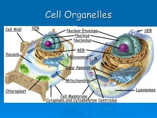
Organelles of eukaryotic organelles. Animal cell. Plant cell. Organelles and their function. http://www.molecularexpressions.com/cells/index.html http://www.uic.edu/classes/bios/bios100/summer2002/lectures.htm http://www.emc.maricopa.edu/faculty/farabee/BIOBK/BioBookCELL2.html. Cell wall.

E N D
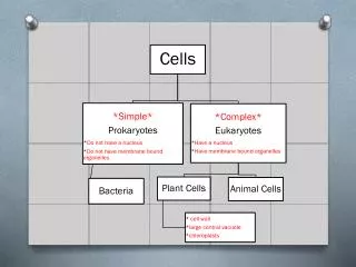
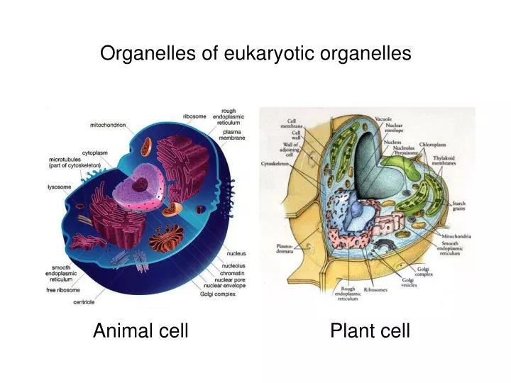
Organelles of eukaryotic organelles Animal cell Plant cell
Organelles and their function http://www.molecularexpressions.com/cells/index.html http://www.uic.edu/classes/bios/bios100/summer2002/lectures.htm http://www.emc.maricopa.edu/faculty/farabee/BIOBK/BioBookCELL2.html
Cell wall The cell wall mainly distinguishes plant cells from animal cells. Structure: This cell wall is generally made up of polysaccharides. Plant cell walls usually contain cellulose. Cellulose provides a stiff and rigid environment for the cell to live in. Function: The cell wall provides the protoplast, or living cells, with mechanical protection and a chemically buffered environment. This function allows the cell to perform homeostatis upon itself, and live in a natural and contained environment. The cell wall is a wall that allows for the circulation and distribution of water, minerals, and other small nutrient molecules into and out of the cell. It provides rigid building blocks from which stable structures such as leaves and stems can be produced. This gives the cell a stable area and self contianed environment. Lastly, it provides a storage site of regulatory molecules that sense the presence of pathogenic microbes and control the development of tissues within the cell.
Plant cell wall Plant cells are surrounded by a rigid, semi-permeable cell wall. The cell wall is comprised mainly polysaccharides with some proteins and lipids. The three main polysaccharide components of the cell wall are cellulose, an unbranched polymers of β-(1-4)-D-glyco-pyranosyl units associated in microfibril bundles. The microfibrils are cross-linked by hemicellulose (a branched polymer of β-(1-4)-D-xylo-pyranosyl units). This cross-linked structure is embedded in a matrix of pectin (primarily containing an α-(1-4) poly-galacturonic acid backbone which can be randomly acetylated and methylated.
Plant cell wall • The main chemical components of the primary plant cell wall include cellulose (in the form of organized microfibrils; see Figure 1), a complex carbohydrate made up of several thousand glucose molecules linked end to end. In addition, the cell wall contains two groups of branched polysaccharides, the pectins and cross-linking glycans. Organized into a network with the cellulose microfibrils, the cross-linking glycans increase the tensile strength of the cellulose, whereas the coextensive network of pectins provides the cell wall with the ability to resist compression. In addition to these networks, a small amount of protein can be found in all plant primary cell walls. Some of this protein is thought to increase mechanical strength and part of it consists of enzymes, which initiate reactions that form, remodel, or breakdown the structural networks of the wall. Such changes in the cell wall directed by enzymes are particularly important for fruit to ripen and leaves to fall in autumn. • The secondary plant cell wall, which is often deposited inside the primary cell wall as a cell matures, sometimes has a composition nearly identical to that of the earlier-developed wall. More commonly, however, additional substances, especially lignin, are found in the secondary wall. Lignin is the general name for a group of polymers of aromatic alcohols that are hard and impart considerable strength to the structure of the secondary wall. Lignin is what provides the favorable characteristics of wood to the fiber cells of woody tissues and is also common in the secondary walls of xylem vessels, which are central in providing structural support to plants. Lignin also makes plant cell walls less vulnerable to attack by fungi or bacteria, as do cutin, suberin, and other waxy materials that are sometimes found in plant cell walls. • A specialized region associated with the cell walls of plants, and sometimes considered an additional component of them, is the middle lamella (see Figure 1). Rich in pectins, the middle lamella is shared by neighboring cells and cements them firmly together. Positioned in such a manner, cells are able to communicate with one another and share their contents through special conduits. Termed plasmodesmata, these small passages penetrate the middle lamella as well as the primary and secondary cell walls, providing pathways for transporting cytoplasmic molecules from one cell to another. http://www.molecularexpressions.com/cells/index.html
Yeast cell wall In yeast, the cell wall comprises ~30 % of the dry weight of the cell. The yeast cell wall is made of ~25% helical β(1-3) and β(1-6)-D-glucans and ~25% oligo-mannans, ~20 % protein, ~10% lipids, and some chitin. The protein component exists pre-dominantly as a mannoprotein complex. Covalent linkages are reported to exist as β(1-4)-linkages between the reducing ends of chitin and the non-reducing end of b(1-3)-glucans as well as among glycoproteins, β(1-6)-glucans, and β(1-3)-glucans.
Plasma membrane • All living cells, prokaryotic and eukaryotic, have a plasma membrane that encloses their contents and serves as a semi-porous barrier to the outside environment. The membrane acts as a boundary, holding the cell constituents together and keeping other substances from entering. The plasma membrane is permeable to specific molecules, however, and allows nutrients and other essential elements to enter the cell and waste materials to leave the cell. Small molecules, such as oxygen, carbon dioxide, and water, are able to pass freely across the membrane, but the passage of larger molecules, such as amino acids and sugars, is carefully regulated. • According to the accepted current theory, known as the fluid mosaic model, the plasma membrane is composed of a double layer (bilayer) of lipids, oily substances found in all cells (see Figure 1). Most of the lipids in the bilayer can be more precisely described as phospholipids, that is, lipids that feature a phosphate group at one end of each molecule. Phospholipids are characteristically hydrophilic ("water-loving") at their phosphate ends and hydrophobic ("water-fearing") along their lipid tail regions. In each layer of a plasma membrane, the hydrophobic lipid tails are oriented inwards and the hydrophilic phosphate groups are aligned so they face outwards, either toward the aqueous cytosol of the cell or the outside environment. Phospholipids tend to spontaneously aggregate by this mechanism whenever they are exposed to water. • Within the phospholipid bilayer of the plasma membrane, many diverse proteins are embedded, while other proteins simply adhere to the surfaces of the bilayer. Some of these proteins, primarily those that are at least partially exposed on the external side of the membrane, have carbohydrates attached to their outer surfaces and are, therefore, referred to as glycoproteins. The positioning of proteins along the plasma membrane is related in part to the organization of the filaments that comprise the cytoskeleton, which help anchor them in place. The arrangement of proteins also involves the hydrophobic and hydrophilic regions found on the surfaces of the proteins: hydrophobic regions associate with the hydrophobic interior of the plasma membrane and hydrophilic regions extend past the surface of the membrane into either the inside of the cell or the outer environment. http://www.molecularexpressions.com/cells/index.html
Plasma membrane proteins • Plasma membrane proteins function in several different ways. Many of the proteins play a role in the selective transport of certain substances across the phospholipid bilayer, either acting as channels or active transport molecules. Others function as receptors, which bind information-providing molecules, such as hormones, and transmit corresponding signals based on the obtained information to the interior of the cell. Membrane proteins may also exhibit enzymatic activity, catalyzing various reactions related to the plasma membrane. • Since the 1970s, the plasma membrane has been frequently described as a fluid mosaic, which is reflective of the discovery that oftentimes the lipid molecules in the bilayer can move about in the plane of the membrane. However, depending upon a number of factors, including the exact composition of the bilayer and temperature, plasma membranes can undergo phase transitions which render their molecules less dynamic and produce a more gel-like or nearly solid state. Cells are able to regulate the fluidity of their plasma membranes to meet their particular needs by synthesizing more of certain types of molecules, such as those with specific kinds of bonds that keep them fluid at lower temperatures. The presence of cholesterol and glycolipids, which are found in most cell membranes, can also affect molecular dynamics and inhibit phase transitions. http://www.molecularexpressions.com/cells/index.html
Mitochondria Mitochondria are found in ALL eukaryotic cells (even in plant cells) They are the site of aerobic respiration sugars + O2 - - > ATP + CO2 + H2O They contain DNA which codes for mitochondrial proteins, ribosomes, etc. They divide by a process similar to binary fission when cell divides They contain a double membrane system Inner Membrane forms the Cristae which are invaginations into interior region. They are the site of energy generation The Matrix is the soluble portion of the mitochondria. It is the site of carbon metabolism, location of mDNA and of mitochondrial protein synthesis
Mitochondria The number of mitochondria present in a cell depends upon the metabolic requirements of that cell, and may range from a single large mitochondrion to thousands of the organelles. Mitochondria, which are found in nearly all eukaryotes, including plants, animals, fungi, and protists, are large enough to be observed with a light microscope and were first discovered in the 1800s. The name of the organelles was coined to reflect the way they looked to the first scientists to observe them, stemming from the Greek words for "thread" and "granule." For many years after their discovery, mitochondria were commonly believed to transmit hereditary information. It was not until the mid-1950s when a method for isolating the organelles intact was developed that the modern understanding of mitochondrial function was worked out. The elaborate structure of a mitochondrion is very important to the functioning of the organelle (see Figure 1). Two specialized membranes encircle each mitochondrion present in a cell, dividing the organelle into a narrow intermembrane space and a much larger internal matrix, each of which contains highly specialized proteins. The outer membrane of a mitochondrion contains many channels formed by the protein porin and acts like a sieve, filtering out molecules that are too big. Similarly, the inner membrane, which is highly convoluted so that a large number of infoldings called cristae are formed, also allows only certain molecules to pass through it and is much more selective than the outer membrane. To make certain that only those materials essential to the matrix are allowed into it, the inner membrane utilizes a group of transport proteins that will only transport the correct molecules. Together, the various compartments of a mitochondrion are able to work in harmony to generate ATP in a complex multi-step process. Mitochondria are generally oblong organelles, which range in size between 1 and 10 micrometers in length, and occur in numbers that directly correlate with the cell's level of metabolic activity. The organelles are quite flexible, however, and time-lapse studies of living cells have demonstrated that mitochondria change shape rapidly and move about in the cell almost constantly. Movements of the organelles appear to be linked in some way to the microtubules present in the cell, and are probably transported along the network with motor proteins. Consequently, mitochondria may be organized into lengthy traveling chains, packed tightly into relatively stable groups, or appear in many other formations based upon the particular needs of the cell and the characteristics of its microtubular network. http://www.molecularexpressions.com/cells/index.html
Mitochondria Presented in Figure 2 is a digital image of the mitochondrial network as seen through a fluorescence optical microscope. The extensive intertwined network is labeled with a synthetic dye named MitoTracker Red (red fluorescence) that localizes in the respiring mitochondria of living cells in culture. The rare twin nuclei in this cell were counterstained with a blue dye (cyan fluorescence) to denote their centralized location in relation to the mitochondrial network. The mitochondrion is different from most other organelles because it has its own circular DNA (similar to the DNA of prokaryotes) and reproduces independently of the cell in which it is found; an apparent case of endosymbiosis. Mitochondrial DNA is localized to the matrix, which also contains enzymes as well as ribosomes for protein synthesis. Many of the critical metabolic steps of cellular respiration are catalyzed by enzymes that are able to diffuse through the mitochondrial matrix. The other proteins involved in respiration, including the enzyme that generates ATP, are embedded within the mitochondrial inner membrane. Infolding of the cristae dramatically increases the surface area available for hosting enzymes responsible for cellular respiration. Mitochondria are similar to plant chloroplasts in that both organelles are able to produce energy and metabolites that are required by the host cell. Mitochondria are the sites of respiration, and generate chemical energy in the form of ATP by metabolizing sugars, fats, and other chemical fuels with the assistance of molecular oxygen. In most animal species, mitochondria appear to be primarily inherited through the maternal lineage, though some recent evidence suggests that in rare instances mitochondria may also be inherited via a paternal route. Therefore, unlike nuclear DNA, mitochondrial DNA does not get shuffled every generation, so it is presumed to change at a slower rate, which is useful for the study of human evolution. Mitochondrial DNA is also used in forensic science as a tool for identifying corpses or body parts, and has been implicated in a number of genetic diseases, such as Alzheimer's disease and diabetes. http://www.molecularexpressions.com/cells/index.html
Chloroplasts • Found only in plant cells • Contains membrane-bound photosynthetic pigments • Site of photosynthesis and conversion of solar energy to chemical energy in the form of ATP and sugars • Site of CO2 fixation • Site of O2 generation • Site of sugar synthesis (carbon metabolism) • Contain cpDNA which codes for chloroplast proteins, ribosomes, etc. • Site of chloroplast protein synthesis • Divide when plant cell divides • Enclosed in a double membrane envelope that does not invaginate into the chloroplast • Thylakoid is a third internal membrane system • Stroma is soluble portion of chloroplast
Chloroplasts • Chloroplasts are one of the most widely recognized and important characteristics of plants is their ability to conduct photosynthesis, in effect, to make their own food by converting light energy into chemical energy. This process occurs in almost all plant species and is carried out in specialized organelles known as chloroplasts. All of the green structures in plants, including stems and unripened fruit, contain chloroplasts, but the majority of photosynthesis activity in most plants occurs in the leaves. • Chloroplasts are one of several different types of plastids, plant cell organelles that are involved in energy storage and the synthesis of metabolic materials. The colorless leucoplasts, for instance, are involved in the synthesis of starch, oils, and proteins. Yellow-to-red colored chromoplasts manufacture carotenoids, and the green colored chloroplasts contain the pigments chlorophyll a and chlorophyll b, which are able to absorb the light energy needed for photosynthesis to occur. All plastids develop from tiny organelles found in the immature cells of plant meristems (undifferentiated plant tissue) termed proplastids, and those of a particular plant species all contain copies of the same circular genome. • The chloroplast is enclosed in a double membrane and the area between the two layers that make up the membrane is called the intermembrane space. The outer layer of the double membrane is much more permeable than the inner layer, which features a number of embedded membrane transport proteins. Enclosed by the chloroplast membrane is the stroma, a semi-fluid material that contains dissolved enzymes and comprises most of the chloroplast's volume. Since, like mitochondria, chloroplasts possess their own genomes (DNA), the stroma contains chloroplast DNA and special ribosomes and RNAs as well. In higher plants, lamellae, internal membranes with stacks (each termed a granum) of closed hollow disks called thylakoids, are also usually dispersed throughout the stroma. • Light travels as packets of energy called photons and is absorbed in this form by light-absorbing chlorophyll molecules embedded in the thylakoid disks. When these chlorophyll molecules absorb the photons, they emit electrons, which they obtain from water (a process that results in the release of oxygen as a byproduct). The movement of the electrons causes hydrogen ions to be propelled across the membrane surrounding the thylakoid stack, which consequently initiates the formation of an electrochemical gradient that drives the stroma's production of adenosine triphosphate (ATP). In the stroma, the light-independent reactions of photosynthesis, which involve carbon fixation, occur, and low-energy carbon dioxide is transformed into a high-energy compound like glucose. • Plant cells are remarkable in that they have two organelles specialized for energy production: chloroplasts, which create energy via photosynthesis, and mitochondria, which generate energy through respiration, a particularly important process when light is unavailable. Like the mitochondrion, the chloroplast is different from most other organelles because it has its own DNA and reproduces independently of the cell in which it is found; an apparent case of endosymbiosis. http://www.molecularexpressions.com/cells/index.html
Endoplasmic reticulum (ER) The Endoplasmic Reticulum (ER) is an extinsive membranous network continuous with the outer nuclear membrane. The Rough ER has ribosomes and is involved in the synthesis of secretory proteins. The Smooth ER lacks ribosomes and is involved in membrane lipid synthesis.
Endoplasmic reticulum (ER) • The endoplasmic reticulum (ER) is a network of flattened sacs and branching tubules that extends throughout the cytoplasm in plant and animal cells. These sacs and tubules are all interconnected by a single continuous membrane so that the organelle has only one large, highly convoluted and complexly arranged lumen (internal space). Usually referred to as the endoplasmic reticulum cisternal space, the lumen of the organelle often takes up more than 10 percent of the total volume of a cell. The endoplasmic reticulum membrane allows molecules to be selectively transferred between the lumen and the cytoplasm, and since it is connected to the double-layered nuclear envelope, it further provides a pipeline between the nucleus and the cytoplasm. • The endoplasmic reticulum manufactures, processes, and transports a wide variety of biochemical compounds for use inside and outside of the cell. Consequently, many of the proteins found in the cisternal space of the endoplasmic reticulum lumen are there only transiently as they pass on their way to other locations. Other proteins, however, are targeted to constantly remain in the lumen and are known as endoplasmic reticulum resident proteins. These special proteins, which are necessary for the endoplasmic reticulum to carry out its normal functions, contain a specialized retention signal consisting of a specific sequence of amino acids that enables them to be retained by the organelle. • There are two basic kinds of endoplasmic reticulum morphologies: rough and smooth. The surface of rough endoplasmic reticulum is covered with ribosomes, giving it a bumpy appearance when viewed through the microscope. This type of endoplasmic reticulum is involved mainly with the production and processing of proteins that will be exported, or secreted, from the cell. The ribosomes assemble amino acids into protein units, which are transported into the rough endoplasmic reticulum for further processing. • Most proteins exported from the endoplasmic reticulum exit the organelle in vesicles budded from the smooth portion, which has a more even appearance than rough endoplasmic reticulum when viewed through the electron microscope because of the lack of ribosomes. The smooth endoplasmic reticulum in most cells is much less extensive than the rough endoplasmic reticulum and is sometimes alternatively termed transitional. Smooth endoplasmic reticulum is chiefly involved, however, with the production of lipids (fats), building blocks for carbohydrate metabolism, and the detoxification of drugs and poisons. Therefore, in some specialized cells, such as those that are occupied chiefly in lipid and carbohydrate metabolism (brain and muscle) or detoxification (liver), the smooth endoplasmic reticulum is much more extensive and is crucial to cellular function. Smooth endoplasmic reticulum also plays a role in various cellular activities through its storage of calcium and involvement in calcium metabolism. In muscle cells, smooth endoplasmic reticulum releases calcium to trigger muscle contractions... http://www.molecularexpressions.com/cells/index.html
Ribosomes • The "factories" of the cell - involved in protein synthesis • Facilitate the specific coupling of tRNA anticodons with mRNA codons during protein synthesis • May either be free or bound to ER • Made up of two subunits, the large and the small subunit • Both subunits are constructed out of protein and RNA (called rRNA) • The ribosomes of prokaryotes and eukaryotes vary slightly with regard to size and shape
Ribosomes • Ribosomes are composed of approximately 60 percent ribosomal RNA (rRNA) and 40 percent protein. However, though they are generally described as organelles, it is important to note that ribosomes are not bound by a membrane and are much smaller than other organelles. Some cell types may hold a few million ribosomes, but several thousand is more typical. • Ribosomes are mainly found bound to the endoplasmic reticulum and the nuclear envelope, as well as freely scattered throughout the cytoplasm, depending upon whether the cell is plant, animal, or bacteria. The organelles serve as the protein production machinery for the cell and are consequently most abundant in cells that are active in protein synthesis, such as pancreas and brain cells. Some of the proteins synthesized by ribosomes are for the cell's own internal use, especially those that are produced by free ribosomes. Many of the proteins produced by bound ribosomes, however, are transported outside of the cell. • Eukaryote ribosomes are produced and assembled in the nucleolus. Ribosomal proteins enter the nucleolus and combine with the four rRNA strands to create the two ribosomal subunits (one small and one large) that will make up the completed ribosome (see Figure 1). The ribosome units leave the nucleus through the nuclear pores and unite once in the cytoplasm for the purpose of protein synthesis. • The units of a ribosome are often described by their Svedberg (s) values, which are based upon their rate of sedimentation in a centrifuge. The ribosomes in a eukaryotic cell generally have a Svedberg value of 80S and are comprised of 40s and 60s subunits. Prokaryotic cells, on the other hand, contain 70S ribosomes, each of which consists of a 30s and a 50s subunit. As demonstrated by these values, Svedberg units are not additive, so the values of the two subunits of a ribosome do not add up to the Svedberg value of the entire organelle. This is because the rate of sedimentation of a molecule depends upon its size and shape, rather than simply its molecular weight. • Protein synthesis requires the assistance of two other kinds of RNA molecules in addition to rRNA. Messenger RNA (mRNA) provides the template of instructions from the cellular DNA for building a specific protein. Transfer RNA (tRNA) brings the protein building blocks, amino acids, to the ribosome. There are three adjacent tRNA binding sites on a ribosome: the aminoacyl binding site for a tRNA molecule attached to the next amino acid in the protein (as illustrated in Figure 1), the peptidyl binding site for the central tRNA molecule containing the growing peptide chain, and an exit binding site to discharge used tRNA molecules from the ribosome. A single ribosome in a eukaryotic cell can add 2 amino acids to a protein chain every second. In prokaryotes, ribosomes can work even faster, adding about 20 amino acids to a polypeptide every second. • In addition to the most familiar cellular locations of ribosomes, the organelles can also be found inside mitochondria and the chloroplasts of plants. These ribosomes notably differ in size and makeup than other ribosomes found in eukaryotic cells, and are more akin to those present in bacteria and blue-green algae cells. The similarity of mitochondrial and chloroplast ribosomes to prokaryotic ribosomes is generally considered strong supportive evidence that mitochondria and chloroplasts evolved from ancestral prokaryotes. http://www.molecularexpressions.com/cells/index.html
Golgi • Flattened vesicles in stacks which receive protein from ER • Form secretory vesicles to transport proteins to different parts of the cell (vacuole, lysosome, etc) or for secretion • cis face - "receiving" side of Golgi apparatus • trans face - "shipping" side of Golgi apparatus
Golgi • The Golgi apparatus (GA), also called Golgi body or Golgi complex and found universally in both plant and animal cells, is typically comprised of a series of five to eight cup-shaped, membrane-covered sacs called cisternae that look something like a stack of deflated balloons. In some unicellular flagellates, however, as many as 60 cisternae may combine to make up the Golgi apparatus. Similarly, the number of Golgi bodies in a cell varies according to its function. Animal cells generally contain between ten and twenty Golgi stacks per cell, which are linked into a single complex by tubular connections between cisternae. • The Golgi apparatus is often considered the distribution and shipping department for the cell's chemical products. It modifies proteins and lipids (fats) that have been built in the endoplasmic reticulum and prepares them for export outside of the cell or for transport to other locations in the cell. Proteins and lipids built in the smooth and rough endoplasmic reticulum bud off in tiny bubble-like vesicles that move through the cytoplasm until they reach the Golgi complex. The vesicles fuse with the Golgi membranes and release their internally stored molecules into the organelle. Once inside, the compounds are further processed by the Golgi apparatus, which adds molecules or chops tiny pieces off the ends. When completed, the product is extruded from the GA in a vesicle and directed to its final destination inside or outside the cell. The exported products are secretions of proteins or glycoproteins that are part of the cell's function in the organism. Other products are returned to the endoplasmic reticulum or may undergo maturation to become lysosomes. • The modifications to molecules that take place in the Golgi apparatus occur in an orderly fashion. Each Golgi stack has two distinct ends, or faces. The cis face of a Golgi stack is the end of the organelle where substances enter from the endoplasmic reticulum for processing, while the trans face is where they exit in the form of smaller detached vesicles. Consequently, the cis face is found near the endoplasmic reticulum, from whence most of the material it receives comes, and the trans face is positioned near the plasma membrane of the cell, to where many of the substances it modifies are shipped. The chemical make-up of each face is different and the enzymes contained in the lumens (inner open spaces) of the cisternae between the faces are distinctive. • Proteins, carbohydrates, phospholipids, and other molecules formed in the endoplasmic reticulum are transported to the Golgi apparatus to be biochemically modified during their transition from the cis to the trans poles of the complex. Enzymes present in the Golgi lumen modify the carbohydrate (or sugar) portion of glycoproteins by adding or subtracting individual sugar monomers. In addition, the Golgi apparatus manufactures a variety of macromolecules on its own, including a variety of polysaccharides. The Golgi complex in plant cells produces pectins and other polysaccharides specifically needed by for plant structure and metabolism. http://www.molecularexpressions.com/cells/index.html
Peroxisomes • Peroxisomes/Microbodies are a diverse group of organelles that are found in the cytoplasm of almost all cells, roughly spherical, and bound by a single membrane. There are several types of microbodies, but peroxisomes are the most common. All eukaryotes are comprised of one or more cells that contain peroxisomes. The organelles were first discovered by the Belgian scientist Christian de Duve, who also discovered lysosomes.
Peroxisomes • Peroxisomes contain a variety of enzymes, which primarily function together to rid the cell of toxic substances, and in particular, hydrogen peroxide (a common byproduct of cellular metabolism). These organelles contain enzymes that convert the hydrogen peroxide to water, rendering the potentially toxic substance safe for release back into the cell. Some types of peroxisomes, such as those in liver cells, detoxify alcohol and other harmful compounds by transferring hydrogen from the poisons to molecules of oxygen (a process termed oxidation). Others are more important for their ability to initiate the production of phospholipids, which are typically used in the formation of membranes. • Peroxisomes self-replicate, but unlike self-replicating mitochondria peroxisomes do not have their own internal DNA. Consequently, the organelles must import the proteins they need to make copies of themselves from the surrounding cytosol. The importation process of peroxisomes is not yet well understood, but it appears to be heavily dependent upon peroxisomal targeting signals composed of specific amino acid sequences. These signals are thought to interact with receptor proteins present in the cytosol and docking proteins present in the peroxisomal membrane. • Since the early 1980s, a number of metabolic disorders have been discovered to be caused by molecular defects in peroxisomes. Two major categories have been described so far. The first category consists of disorders of peroxisome biogenesis in which the organelle fails to develop normally, causing defects in numerous peroxisomal proteins. The second category involves defects of single peroxisomal enzymes. Studies indicate that approximately one in every 20,000 people has some type of a peroxisomal disorder. The most serious of these disorders is Zellweger syndrome, which is characterized by an absence or reduced number of peroxisomes in the cells. Present in patients at birth (congenital), Zellweger syndrome has no cure or effective treatment and usually causes death within the first year of life. http://www.molecularexpressions.com/cells/index.html
Lysosomes • The main function of lysosomes is digestion. Lysosomes break down cellular waste products and debris from outside the cell into simple compounds, which are transferred to the cytoplasm as new cell-building materials. They are involved in processes like endocytosis, phagocytosis and autophagy.
Lysosomes • The main function of these microscopic organelles is to serve as digestion compartments for cellular materials that have exceeded their lifetime or are otherwise no longer useful. In this regard, the lysosomes recycle the cell's organic material in a process known as autophagy. Lysosomes break down cellular waste products, fats, carbohydrates, proteins, and other macromolecules into simple compounds, which are then transferred back into the cytoplasm as new cell-building materials. To accomplish the tasks associated with digestion, the lysosomes utilize about 40 different types of hydrolytic enzymes, all of which are manufactured in the endoplasmic reticulum and modified in the Golgi apparatus. Lysosomes are often budded from the membrane of the Golgi apparatus, but in some cases they develop gradually from late endosomes, which are vesicles that carry materials brought into the cell by a process known as endocytosis. • Like other microbodies, lysosomes are spherical organelles contained by a single layer membrane, though their size and shape varies to some extent. This membrane protects the rest of the cell from the harsh digestive enzymes contained in the lysosomes, which would otherwise cause significant damage. The cell is further safeguarded from exposure to the biochemical catalysts present in lysosomes by their dependency on an acidic environment. With an average pH of about 4.8, the lysosomal matrix is favorable for enzymatic activity, but the neutral environment of the cytosol renders most of the digestive enzymes inoperative, so even if a lysosome is ruptured, the cell as a whole may remain uninjured. The acidity of the lysosome is maintained with the help of hydrogen ion pumps, and the organelle avoids self-digestion by glucosylation of inner membrane proteins to prevent their degradation. • Lysosomes are found in all animal cells, but are most numerous in disease-fighting cells, such as white blood cells. This is because white blood cells must digest more material than most other types of cells in their quest to battle bacteria, viruses, and other foreign intruders. Several human diseases are caused by lysosome enzyme disorders that interfere with cellular digestion. Tay-Sachs disease, for example, is caused by a genetic defect that prevents the formation of an essential enzyme that breaks down complex lipids called gangliosides. Accumulation of these lipids damages the nervous system, causes mental retardation, and death in early childhood. Also, arthritis inflammation and pain are related to the escape of lysosome enzymes. http://www.molecularexpressions.com/cells/index.html
Vacuole • The Plant Central Vacuole is a major storage space in center of plant cell with many functions • Digestive - break down of macromolecules • Storage - ions, sugars, amino acids, toxic waste • Maintaining cell rigidity - high ionic concentration generates high water potential
Vacuole • Among its roles in plant cell function, the central vacuole stores salts, minerals, nutrients, proteins, pigments, helps in plant growth, and plays an important structural role for the plant. Under optimal conditions, the vacuoles are filled with water to the point that they exert a significant pressure against the cell wall. This helps maintain the structural integrity of the plant, along with the support from the cell wall, and enables the plant cell to grow much larger without having to synthesize new cytoplasm. In most cases, the plant cytoplasm is confined to a thin layer positioned between the plasma membrane and the tonoplast, yielding a large ratio of membrane surface to cytoplasm. • The structural importance of the plant vacuole is related to its ability to control turgor pressure. Turgor pressure dictates the rigidity of the cell and is associated with the difference between the osmotic pressure inside and outside of the cell. Osmotic pressure is the pressure required to prevent fluid diffusing through a semipermeable membrane separating two solutions containing different concentrations of solute molecules. • Plant vacuoles are also important for their role in molecular degradation and storage. Sometimes these functions are carried out by different vacuoles in the same cell, one serving as a compartment for breaking down materials (similar to the lysosomes found in animal cells), and another storing nutrients, waste products, or other substances. Several of the materials commonly stored in plant vacuoles have been found to be useful for humans, such as opium, rubber, and garlic flavoring, and are frequently harvested. Vacuoles also often store the pigments that give certain flowers their colors, which aid them in the attraction of bees and other pollinators, but also can release molecules that are poisonous, odoriferous, or unpalatable to various insects and animals, thus discouraging them from consuming the plant. • Vacuoles are membrane-bound sacs within the cytoplasm of a cell that function in several different ways. In mature plant cells, vacuoles tend to be very large and are extremely important in providing structural support, as well as serving functions such as storage, waste disposal, protection, and growth. Many plant cells have a large, singlecentral vacuole that typically takes up most of the room in the cell (80 percent or more). • The central vacuole in plant cells (see Figure 1) is enclosed by a membrane termed the tonoplast, an important and highly integrated component of the plant internal membrane network (endomembrane) system. This large vacuole slowly develops as the cell matures by fusion of smaller vacuoles derived from the endoplasmic reticulum and Golgi apparatus. Because the central vacuole is highly selective in transporting materials through its membrane, the chemical palette of the vacuole solution (termed the cell sap) differs markedly from that of the surrounding cytoplasm. For instance, some vacuoles contain pigments that give certain flowers their characteristic colors. The central vacuole also contains plant wastes that taste bitter to insects and animals, while developing seed cells use the central vacuole as a repository for protein storage. http://www.molecularexpressions.com/cells/index.html
Centrioles • Centrioles are found only in animal cells. • These paired organelles are typically located together near the nucleus in the centrosome, a granular mass that serves as an organizing center for micro-tubules. Within the centrosome, the centrioles are positioned so that they are at right angles to each other, as illustrated in Figure 1. Each centriole is made of nine bundles of microtubules (three per bundle) arranged in a ring.
Centrioles • Centrioles play a notable role in cell division. During interphase of an animal cell, the centrioles and other components of the centrosome are duplicated, though scientists are not yet sure how this duplication takes place. At first the two pairs of centrioles remain in close proximity to each other, but as mitosis initiates, the original centrosome divides and the pairs are split up so that one set of centrioles is located in each of the new microtubule-organizing centers. These new centers radiate microtubules in star-shaped clusters known as asters. As the asters move to opposing poles of the cells, the microtubules, with the help of the centrioles, become organized into a spindle-shaped formation that spans the cell (see Figure 2). These spindle fibers act as guides for the alignment of the chromosomes as they separate later during the process of cell division. • Though centrioles play a role in the mitosis of animal cells, plant cells are able to reproduce without them. Researchers have, therefore, been very interested in determining exactly how important the organelles really are. Studies have shown that certain animal cells, particularly female gametes (oocytes), can successfully divide even when their centrioles are destroyed. Some investigators have also found, however, that the absence of centrioles in animal cells is associated with an increased number of divisional errors and substantial delays in the mitotic process, especially before chromosome segregation. Consequently, it has been suggested that centrioles evolved as a refinement of the cell, making mitosis a much more efficient and less error-prone process. http://www.molecularexpressions.com/cells/index.html
Nucleus • The Nucleus contains a double membrane with pores • The outer nuclear membrane is continuous with the ER • The nuclear matrix contains a protein fibrilar network • The nucleoplasm is the fluid substance in which the solutes of the nucleus are dissolved • Chromosomes are complexes consisting of protein and DNA. They contain the genetic information of a cell • The nucleolus is a specific region of the nucleus involved in the synthesis and assembly of ribosomes
Nucleus • The nucleus is a highly specialized organelle that serves as the information processing and administrative center of the cell. This organelle has two major functions: it stores the cell's hereditary material, or DNA, and it coordinates the cell's activities, which include growth, intermediary metabolism, protein synthesis, and reproduction (cell division). • Only the cells of advanced organisms, known as eukaryotes, have a nucleus. Generally there is only one nucleus per cell, but there are exceptions, such as the cells of slime molds and the Siphonales group of algae. Simpler one-celled organisms (prokaryotes), like the bacteria and cyanobacteria, don't have a nucleus. In these organisms, all of the cell's information and administrative functions are dispersed throughout the cytoplasm. • The spherical nucleus typically occupies about 10 percent of a eukaryotic cell's volume, making it one of the cell's most prominent features. A double-layered membrane, the nuclear envelope, separates the contents of the nucleus from the cellular cytoplasm. The envelope is riddled with holes called nuclear pores that allow specific types and sizes of molecules to pass back and forth between the nucleus and the cytoplasm. It is also attached to a network of tubules and sacs, called the endoplasmic reticulum, where protein synthesis occurs, and is usually studded with ribosomes (see Figure 1). • The semifluid matrix found inside the nucleus is called nucleoplasm. Within the nucleoplasm, most of the nuclear material consists of chromatin, the less condensed form of the cell's DNA that organizes to form chromosomes during mitosis or cell division. The nucleus also contains one or more nucleoli, organelles that synthesize protein-producing macromolecular assemblies called ribosomes, and a variety of other smaller components, such as Cajal bodies, GEMS (Gemini of coiled bodies), and interchromatin granule clusters. • Chromatin and Chromosomes - Packed inside the nucleus of every human cell is nearly 6 feet of DNA, which is divided into 46 individual molecules, one for each chromosome and each about 1.5 inches long. Packing all this material into a microscopic cell nucleus is an extraordinary feat of packaging. For DNA to function, it can't be crammed into the nucleus like a ball of string. Instead, it is combined with proteins and organized into a precise, compact structure, a dense string-like fiber called chromatin. • The Nucleolus - The nucleolus is a membrane-less organelle within the nucleus that manufactures ribosomes, the cell's protein-producing structures. Through the microscope, the nucleolus looks like a large dark spot within the nucleus. A nucleus may contain up to four nucleoli, but within each species the number of nucleoli is fixed. After a cell divides, a nucleolus is formed when chromosomes are brought together into nucleolar organizing regions. During cell division, the nucleolus disappears. Some studies suggest that the nucleolus may be involved with cellular aging and, therefore, may affect the senescence of an organism. http://www.molecularexpressions.com/cells/index.html
Nuclear pores • The Nuclear Envelope - The nuclear envelope is a double-layered membrane that encloses the contents of the nucleus during most of the cell's lifecycle. The space between the layers is called the perinuclear space and appears to connect with the rough endoplasmic reticulum. The envelope is perforated with tiny holes called nuclear pores. These pores regulate the passage of molecules between the nucleus and cytoplasm, permitting some to pass through the membrane, but not others. The inner surface has a protein lining called the nuclear lamina, which binds to chromatin and other nuclear components. During mitosis, or cell division, the nuclear envelope disintegrates, but reforms as the two cells complete their formation and the chromatin begins to unravel and disperse. • Nuclear Pores - The nuclear envelope is perforated with holes called nuclear pores. These pores regulate the passage of molecules between the nucleus and cytoplasm, permitting some to pass through the membrane, but not others. Building blocks for DNA and RNA are allowed into the nucleus as well as molecules that provide the energy for constructing genetic material. http://www.molecularexpressions.com/cells/index.html
Cytoskeleton • Cytoskeleton - flexible tubular scaffold of microfilaments • maintains cell shape and provides support • anchors organelles & enzymes to specific regions of the cell • contractility and movement (amoeboid movement) • intracellular transport - tracks for vesicle and organelle movement by motor proteins • Cytoskeleton components • Microfilaments • solid protein (actin) which is assembled at one end and disassembled at the other end • Intermediate filaments - rope-like fibrous proteins • provide structural reinforcement • anchor organelles • keep nucleus in place • Microtubules - hollow tubes of tubulin (a globular protein) • maintains cell shape • anchor organelles • movement of organelles • track for motor proteins
Cytoskeleton: Microfilaments • Common to all eukaryotic cells, these filaments are primarily structural in function and are an important component of the cytoskeleton, along with microtubules and often the intermediate filaments. Microfilaments range from 5 to 9 nanometers in diameter and are designed to bear large amounts of tension. In association with myosin, microfilaments help to generate the forces used in cellular contraction and basic cell movements. The filaments also enable a dividing cell to pinch off into two cells and are involved in amoeboid movements of certain types of cells. • Microfilaments are solid rods made of a protein known as actin. When it is first produced by the cell, actin appears in a globular form (G-actin; see Figure 1). In microfilaments (actin filaments) long polymerized chains of the molecules are intertwined in a helix, creating a filamentous form of the protein (F-actin). All of the subunits that compose a microfilament are connected in such a way that they have the same orientation. Due to this fact, each microfilament exhibits polarity, the two ends of the filament being distinctly different. This polarity affects the growth rate of microfilaments, one end (termed the plus end) typically assembling and disassembling faster than the other (the minus end). • Unlike microtubules, which typically extend out from the centrosome of a cell, microfilaments are typically nucleated at the plasma membrane. Therefore, the periphery (edges) of a cell generally contains the highest concentration of microfilaments. A number of external factors and a group of special proteins influence microfilament characteristics, however, and enable them to make rapid changes if needed, even if the filaments must be completely disassembled in one region of the cell and reassembled somewhere else. When found directly beneath the plasma membrane, microfilaments are considered part of the cell cortex, which regulates the shape and movement of the cell's surface. Consequently, microfilaments play a key role in development of various cell surface projections (as illustrated in Figure 2). • Illustrated in Figure 2 is a fluorescence digital image of a skin fibroblast cell stained with fluorescent probes targeting the nucleus (blue) and the actin cytoskeletal network (green). http://www.molecularexpressions.com/cells/index.html
Cytoskeleton: Microtubules • These straight, hollow cylinders are found throughout the cytoplasm of all eukaryotic cells (prokaryotes don't have them) and carry out a variety of functions, ranging from transport to structural support. Microtubules, which are about 25 nanometers in diameter, form part of the cytoskeleton that gives structure and shape to a cell, and also serve as conveyor belts moving other organelles throughout the cytoplasm. In addition, microtubules are the major components of cilia and flagella, and participate in the formation of spindle fibers during cell division (mitosis). The length of microtubules in the cell varies between 200 nanometers and 25 micrometers, depending upon the task of a particular microtubule and the state of the cell's life cycle. • Microtubules are biopolymers that are composed of subunits made from an abundant globular cytoplasmic protein known as tubulin, as illustrated in Figure 1. Each subunit of the microtubule is made of two slightly different but closely related simpler units called alpha-tubulin and beta-tubulin that are bound very tightly together to form heterodimers. In a microtubule, the subunits are organized in such a way that they all point the same direction to form 13 parallel protofilaments. This organization gives the structure polarity, with only the alpha-tubulin proteins exposed at one end and only beta-tubulin proteins at the other. • By adding or removing globular tubulin proteins, the length of polymeric microtubules can be increased or decreased. Because the two ends of a microtubule are not the same, however, the rate at which growth or depolymerization occurs at each pole is different. The end of a polarized filament that grows and shrinks the fastest is known as the plus end and the opposing end is called the minus end. • Presented in Figure 2 is a digital image of the microtubule network as seen through a fluorescence optical microscope. The extensive intertwined network is labeled with primary antibodies to alpha-tubulin, which are then stained with secondary antibodies containing a green fluorescent dye. • In addition to their structural support role, microtubules also serve as a highway system along which organelles can be transported with the aid of motor proteins. For instance, the microtubule network interconnects the Golgi apparatus with the plasma membrane to guide secretory vesicles for export, and also transports mitochondria back and forth in the cytoplasm.The motor proteins involved in organelle transport operate by altering their three-dimensional conformation using adenosine triphosphate (ATP) as fuel to move back and forth along a microtubule. http://www.molecularexpressions.com/cells/index.html
Cytoskeleton: Intermediate filaments • Intermediate filaments are a broad class of fibrous proteins that play an important role as both structural and functional elements of the cytoskeleton. Ranging in size from 8 to 12 nm (in diameter; see Figure 1), intermediate filaments function as tension-bearing elements to help maintain cell shape and rigidity, and serve to anchor in place several organelles. Intermediate filaments are also involved in formation of the nuclear lamina, a net-like meshwork array that lines the inner nuclear membrane and governs the shape of the nucleus. • Although all eukaryotes contain the common cytoskeletal elements actin and tubulin (both free in the cytoplasm and polymerized in the form of microfilaments and microtubules), intermediate filaments are found only in some metazoan species, including vertebrates. In vertebrates, intermediate filament presence and composition varies with the tissue type. Examples are keratins, vimentin and desmin, peripherin, neurofilaments, and glial fibrillary acidic protein (GFAP). Associated proteins bind to intermediate filaments, either to improve stability (through crosslinking) or to provide attachment sites for other protein assemblies, such as actin filaments and microtubules. • Another highly specialized class of intermediate filaments are the nuclearlamins, which constitute the fibrous protein network that lines the inside of the nuclear membrane. Due to their close proximity to the membrane, nuclear lamins help attach the chromosomes to the nuclear membrane and provide anchorage points for the nuclear pores. • As illustrated in Figure 1, intermediate filament monomer peptides are an elongated fibrous class of proteins with a central alpha-helical region capped with globular ends at both the amino and carboxylic acid termini. Two of the monomer units form a coiled-coil dimer that self-associates in an anti-parallel arrangement to form a staggered tetramer, which is the analogous soluble subunit for the globular actin monomer and the tubulin heterodimer (existing free in the cytoplasm). Tetramer units pack together laterally to form a sheet of eight parallel protofilaments that are supercoiled into a tight bundle. Each tightly coiled intermediate filament cross section reveals 32 individual alpha-helical peptides, which renders the filament easy to bend but quite difficult to break, thus accounting for the extreme structural rigidity. • Mutations in intermediate filament genes lead to a host of rather uncommon diseases. For example, defective keratins in skin tissue lead to a disorder known as epidermolysis bullosa simplex, manifested by skin blisters produced with even a slight mechanical stress. Several neurodegenerative diseases, such as amyotrophic lateral sclerosis (ALS or Lou Gehrig's Disease), are associated with malfunctions in the intermediate filament (neurofilament) network, and defects in desmin intermediate filaments produce muscle disorders. http://www.molecularexpressions.com/cells/index.html
Cytoplasm/Cytosol • The cytoplasm is the site where most cellular activities are done. The cytosol has enzymes that take molecules and break them down , so that the individual organelles can use them as they need to. The most prominent metabolic pathways in the cytoplasm are glycolysis and fatty acid synthesis. The cytosol also contains the cytoskeleton which gives the cell its shape and can help in the movement of the cell. • Cytoplasm is the watery environment inside the cell that makes up about 70% of the cell. Cytoplasm includes salts, an assortment of organic molecules, including many enzymes that catalyze reactions, as well as water. The cytoplasm is seperated from the wattery extracellular fluid, which is outside, by the plasma membrane.