Download
1 / 41
410 likes | 597 Vues
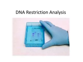
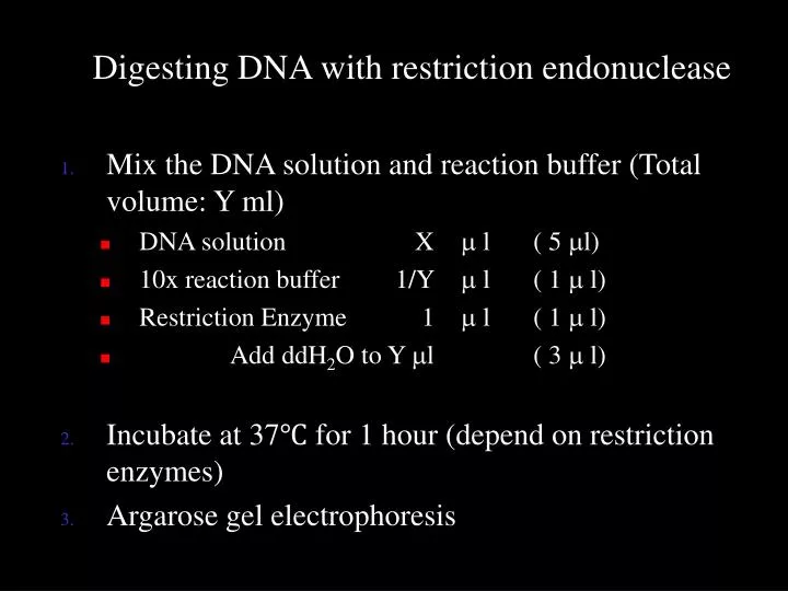
Digesting DNA with restriction endonuclease Mix the DNA solution and reaction buffer (Total volume: Y ml) DNA solution X m l ( 5 m l) 10x reaction buffer 1/Y m l ( 1 m l) Restriction Enzyme 1 m l ( 1 m l) Add ddH 2 O to Y m l ( 3 m l)

E N D
Digesting DNA with restriction endonuclease • Mix the DNA solution and reaction buffer (Total volume: Y ml) • DNA solution X ml ( 5 ml) • 10x reaction buffer 1/Y ml ( 1 ml) • Restriction Enzyme 1 ml ( 1 ml) • Add ddH2O to Y ml ( 3 ml) • Incubate at 37℃ for 1 hour (depend on restriction enzymes) • Argarose gel electrophoresis
Recombinant DNA Technology • Gene cloning or Molecular cloning • The recombinant DNA construction • Transformation • Identification
The recombinantDNA construction Transformation Identification
Restriction Endonucleases • Nomenclature • Eco RI EscherichiacoliR • Hpa I Haemopophilus parainfluenzae • Palindromic sequence • Types of cut ends • 5’ phosphate extension • 3’ hydroxyl extension • Blunt end
Fig 4.2 Palindromic sequence
Isochizomer • Different restriction enzymes recognize different sequences • Cleave within the same target sequences • Sau 3A, Mbo I & Bam HI (5’…GATC…3’) • SalI & XhoI (5’-GTCGAC- & 5’-CTCGAG-)
Ligases • E coli • T4
3’ to pyrimidine 3’ to guanine