Download
1 / 104
1.11k likes | 1.98k Vues
Definition. The term birth injury is used to denote: avoidable and unavoidable mechanical, hypoxic and ischemic injury affecting the infant during labor and delivery. . Birth injuries may result from :Inappropriate or deficient medical skill or attention.They may occur, despite skilled and co

E N D
1. Fetal Birth Injuries Dr. Ashraf Fouda
Domiatte General Hospital
2. Definition The term birth injury is used to denote:
avoidable and unavoidable
mechanical, hypoxic and ischemic injury
affecting the infant
during
labor and delivery.
3. Birth injuries may result from :
Inappropriate or deficient medical skill or attention.
They may occur, despite skilled and competent obstetric care. Definition
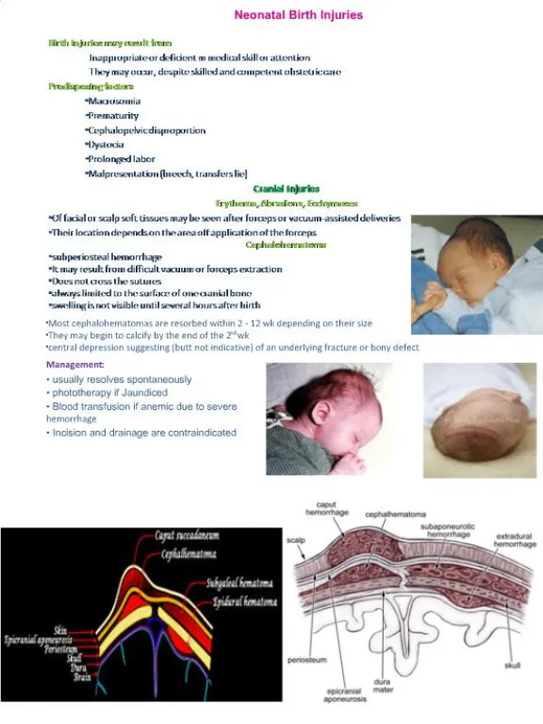
4. Incidence Has been estimated at 2-7/1,000 live births. Predisposing factors:
Macrosomia,
Prematurity,
Cephalopelvic disproportion,
Dystocia,
Prolonged labor, and
Breech presentation.
5. 5-8/100,000 infants die of birth trauma, and
25/100,000 die of anoxic injuries;
Such injuries represent 2-3% of infant deaths. Incidence
6. Cranial Injuries
7. Erythema, abrasions, ecchymoses, Of facial or scalp soft tissues may be seen after forceps or vacuum-assisted deliveries.
Their location depends on the area of application of the forceps.
8. Subconjunctival ,retinal hemorrhages and petechiae of the skin of the head and neck All are common.
All are probably secondary to a sudden increase in intrathoracic pressure during passage of the chest through the birth canal.
Parents should be assured that they are temporary and the result of normal hazards of delivery.
9. Molding Molding of the head and overriding of the parietal bones are frequently associated with caput succedaneum and become more evident after the caput has receded but disappear during the first weeks of life.
Rarely, a hemorrhagic caput may result in shock and require blood transfusion.
10. Caput succedaneum Diffuse, sometimes ecchymotic, edematous swelling of the soft tissues of the scalp involving the portion presenting during vertex delivery.
It may extend across the midline and across suture lines.
The edema disappears within the first few days of life.
11. Analogous swelling, discoloration, and distortion of the face are seen in face presentations.
No specific treatment is needed, but if there are extensive ecchymoses, phototherapy for hyperbilirubinemia may be indicated. Caput succedaneum
12. Cephalhaematoma It is a subperiosteal haematoma most commonly lies over one parietal bone.
It may result from difficult vacuum or forceps extraction .
13. Management:
- It usually resolves spontaneously.
- Vitamin K 1 mg IM is given. Cephalhaematoma
16. Cephalohematoma Is a subperiosteal hemorrhage, so it is always limited to the surface of one cranial bone.
There is no discoloration of the overlying scalp, and swelling is usually not visible until several hours after birth, because subperiosteal bleeding is a slow process.
An underlying skull fracture, usually linear and not depressed, is occasionally associated with cephalohematoma.
17. Cranial meningocele
is differentiated from cephalohematoma by:
Pulsation,
Increased pressure on crying, and the
Radiologic evidence of bony defect.
Most cephalohematomas are resorbed within 2 wk-3 mo, depending on their size.
They may begin to calcify by the end of the 2nd wk. Cephalohematoma
18. A sensation of central depression suggesting( but not indicative )of an underlying fracture or bony defect is
Cephalohematomas
require no treatment, although phototherapy may be necessary to ameliorate hyperbilirubinemia. Cephalohematoma
19. Incision and drainage are contraindicated because of the risk of introducing infection in a benign condition.
A massive cephalohematoma may rarely result in blood loss severe enough to require transfusion.
It may also be associated with a skull fracture, coagulopathy, and intracranial hemorrhage. Cephalohematoma
20. Diagnosis and Differential Diagnosis
21. Fractures of the skull May occur as a result of pressure from :
Forceps or from
The maternal symphysis pubis.
Sacral promontory, or
Ischial spines.
22. Fracture Skull: Usually occurs due to difficult forceps delivery.
It may be:
(1) Vault fracture:
Usually affecting the frontal or parietal bone.
It may be linear or depressed fracture.
It needs no treatment unless there is intracranial haemorrhage.
(2) Fracture base:
Usually associated with intracranial haemorrhage.
23. Linear fractures, the most common, cause no symptoms and require no treatment.
Depressed fractures are usually indentations similar to a dent in a Ping-Pong ball; they usually are a complication of forceps delivery or fetal compression. Fractures of the skull
25. Affected infants may be asymptomatic unless there is associated intracranial injury.
It is advisable to elevate severe depressions to prevent cortical injury from sustained pressure. Fractures of the skull
26. Fracture of the Occipital bone almost causes fatal hemorrhage due to disruption of the underlying vascular sinuses.
It may result during breech deliveries from traction on the hyperextended spine of the infant with the head fixed in the maternal pelvis. Fractures of the skull
28. Intracranial Haemorrhage: Causes:
Sudden compression and decompression of the head as in breech and precipitate labour.
Marked compression by forceps or in cephalopelvic disproportion.
Fracture skull.
29. Predisposing factors:
Prematurity due to physiological hypoprothrombinaemia, fragile blood vessels and liability to trauma.
Asphyxia due to anoxia of the vascular wall .
Blood diseases. Intracranial Haemorrhage:
30. Subdural : results from damage to the superficial veins where the vein of Galen and inferior sagittal sinus combine to form the straight sinus.
Subarachnoid: The vein of Galen is damaged due to tear in the dura at the junction of the falx cerebri and tentorium cerebelli.
Intraventricular :into the brain ventricles.
Intracerebral : into the brain tissues .
In (1) and (2) it is usually due to birth trauma,
in (3) and (4) the foetus is usually a premature exposed to hypoxia. Intracranial Haemorrhage Sites:
31. Clinical picture:
1- Altered consciousness.
2- Flaccidity.
3- Breathing is absent, irregular and periodic or gasping.
4- Eyes: no movement, pupils may be fixed and dilated.
5- Opisthotonus, rigidity, twitches and convulsions.
6- Vomiting .
7- High pitched cry.���
8- Anterior fontanelle is tense and bulging.
9- Lumbar puncture reveals bloody C.S.F. Intracranial Haemorrhage:
32. Investigations:
Ultrasound is of value.
CT scan is the most reliable.
MRI Intracranial Haemorrhage
33. Prophylaxis:
Vitamin K: 10 mg IM to the mother in late pregnancy or early in labour.
Episiotomy: especially in prematures and breech delivery.
Forceps delivery: carried out by an experienced obstetrician respecting the instructions for its use. Intracranial Haemorrhage:
34. Minimal handling, warmth and oxygen to the baby.
No oral feeding for 72 hours.
IV fluids.
Vitamin K 1mg IM.
Lumbar puncture: is diagnostic and therapeutic to relieve the intracranial tension if the anterior fontanelle is bulging.
Sedatives for convulsions.
60 cc. of 10% sodium chloride per rectum to relieve brain oedema.
1 cc of 50% magnesium sulphate IM to relieve brain oedema and convulsions.
Antibiotics : to guard against infections particularly pulmonary. Intracranial Haemorrhage Treatment
35. ETIOLOGY AND EPIDEMIOLOGY Intracranial hemorrhage may result from:
Birth trauma or
Asphyxia and, rarely, from a
Primary hemorrhagic disturbance or
Congenital vascular anomaly.
36. Intracranial hemorrhages often involve the ventricles
( intraventricular hemorrhage [IVH]) of premature infants delivered spontaneously without apparent trauma. ETIOLOGY AND EPIDEMIOLOGY
37. CLINICAL MANIFESTATIONS The incidence of IVH increases with decreasing birthweight:
60-70% of 500- to 750-g infants and
10-20% of 1,000- to 1,500-g infants.
IVH is rarely present at birth; however,
80-90% of cases occur between birth and the 3rd day .
50% occur on the 1st day.
20% to 40% of cases progress during the 1st wk of life.
Delayed hemorrhage may occur in 10-15% of patients after the 1st wk of life.
38. The most common symptoms are:
Diminished or absent Moro reflex.
Poor muscle tone.
Lethargy.
Apnea.
Somnolence. CLINICAL MANIFESTATIONS
39. Periods of apnea,
Pallor, or cyanosis;
Failure to suck well;
Abnormal eye signs;
A high-pitched cry;
Muscular twitches, convulsions, decreased muscle tone, or paralyses;
Metabolic acidosis; shock, and a
Decreased hematocrit or its failure to increase after transfusion may be the first indications.
The fontanel may be tense and bulging. CLINICAL MANIFESTATIONS
40. DIAGNOSIS Intracranial hemorrhage is diagnosed on the basis of the:
History,
Clinical manifestations,
Transfontanel cranial ultrasonography or
Computed tomography (CT), and
41. Lumbar puncture
is indicated in the presence of signs of:
Increased intracranial pressure or
Deteriorating clinical condition
to identify gross subarachnoid hemorrhage or to rule out the possibility of bacterial meningitis DIAGNOSIS
42. PROGNOSIS Neonates with:
( massive hemorrhage associated with tears of the tentorium or falx cerebri)
rapidly deteriorate and may die after birth.
43. PREVENTION The incidence of traumatic intracranial hemorrhage may be reduced by:
judicious management of cephalopelvic disproportion and operative delivery.
44. Fetal or neonatal hemorrhage due to:
Maternal idiopathic thrombocytopenic purpura (ITP) or
Alloimmune thrombocytopenia
may be prevented by maternal treatment with:
Steroids,
Intravenous immunoglobulin, or
Fetal platelet transfusion. PREVENTION
45. The incidence of IVH may be reduced by antenatal steroids and by postnatal administration of low-dose indomethacin.
Vitamin K should be given before delivery to all women receiving phenobarbital or phenytoin during the pregnancy. PREVENTION
46. TREATMENT Seizures are treated with anticonvulsant drugs.
Anemia-shock, requires transfusion with packed red blood cells or fresh frozen plasma.
Acidosis is treated with slow administration of sodium bicarbonate.
47. Symptomatic subdural hemorrhage in large term infants should be treated by removing the subdural fluid collection by means of a spinal needle placed through the lateral margin of the anterior fontanel. TREATMENT
48. Spine and Spinal Cord Strong traction exerted:
When the spine is hyperextended or
When the direction of pull is lateral, or
Forceful longitudinal traction on the trunk while the head is still firmly engaged in the pelvis:
(may produce fracture and separation of the vertebrae).
49. Such injuries, rarely diagnosed clinically, are most likely to occur with shoulder dystocia.
The injury occurs most commonly at the level of the 4th cervical vertebra with cephalic presentations and
The lower cervical-upper thoracic vertebrae with breech presentations. Spine and Spinal Cord
51. Transection of the cord may occur with or without vertebral fractures.
Hemorrhage and edema may produce neurologic signs that are not distinguished from those of transection
(except that they may not be permanent). Spine and Spinal Cord
52. Areflexia,
Loss of sensation, and
Complete paralysis of voluntary motion
Occur below the level of injury Spine and Spinal Cord
53. If the injury is severe, the infant, (who may be in poor condition owing to respiratory depression, shock, or hypothermia),
May deteriorate rapidly to death within several hours before neurologic signs are obvious. Spine and Spinal Cord
54. The course may be protracted, with symptoms and signs appearing at birth or later in the 1st wk; may not be recognized for several days.
Constipation may also be present. Spine and Spinal Cord
55. The diagnosis is confirmed by :
Ultrasonography or MRI.
Treatment of the survivors is:
supportive, including home ventilation; patients often remain permanently injured. Spine and Spinal Cord
56. Peripheral Nerve Injuries
57. Brachial Plexus Palsy: It is due to over traction on
the neck as in:
Shoulder dystocia.�����
After-coming head in breech delivery.
58. Erb's palsy:
It is the common, due to injury to C5 and C6 roots.
The upper limb drops beside the trunk, internally rotated with flexed wrist
(policeman�s or waiter�s tip hand). Brachial Plexus Palsy:
59. (2) Klumpke�s palsy:
It is less common,
Due to injury to C7 and C8 and 1st thoracic roots.
- It leads to paralysis of the muscles of the hand and weakness of the wrist and fingers' flexors. Brachial Plexus Palsy:
60. Treatment
Support to prevent stretching of the paralyzed muscles.
Physiotherapy: massage, exercise and faradic stimulation. Brachial Plexus Palsy:
62. BRACHIAL PALSY Injury to the brachial plexus may cause paralysis of the upper arm with or without paralysis of the forearm or hand or, more commonly, paralysis of the entire arm.
Approximately 45% are associated with shoulder dystocia.
63. These injuries occur in :
Macrosomic infants and when lateral traction is exerted on the head and neck during delivery of the shoulder in a vertex presentation,
When the arms are extended over the head in a breech presentation, or
When excessive traction is placed on the shoulders. BRACHIAL PALSY
65. In Erb-Duchenne paralysis The injury is limited to the 5th and 6th cervical nerves.
The characteristic position consists of:
( Adduction and internal rotation of the arm with pronation of the forearm).
Moro reflex is absent on the affected side
67. There may be some sensory impairment on the outer aspect of the arm.
The power in the forearm and the hand grasp are preserved unless the lower part of the plexus is also injured;
(the presence of the hand grasp is a favorable prognostic sign). In Erb-Duchenne paralysis
68. Klumpke's paralysis Is a rarer form of brachial palsy;
Injury to the 7th and 8th cervical nerves and the 1st thoracic nerve produces a paralyzed hand,
(Horner syndrome)
If the sympathetic fibers of the 1st thoracic root are also injured : paralyzed hand and ipsilateral ptosis and miosis.
69. The mild cases may not be detected immediately after birth.
Differentiation must be made from :
Cerebral injury;
Fracture, dislocation, or epiphyseal separation of the humerus;
Fracture of the clavicle.
MRI demonstrates nerve root rupture or avulsion Klumpke's paralysis
71. The prognosis Depends on whether the nerve was merely injured or was lacerated.
If the paralysis was due to edema and hemorrhage about the nerve fibers, function should return within a few months;
If due to laceration, permanent damage may result.
72. Involvement of the deltoid is usually the most serious problem and may result in a shoulder drop secondary to muscle atrophy.
In general, paralysis of the upper arm has a better prognosis than paralysis of the lower arm. The prognosis
73. Treatment Partial immobilization and appropriate positioning to prevent development of contractures.
In upper arm paralysis: the arm should be abducted, with external rotation at the shoulder and with full supination of the forearm and slight extension at the wrist with the palm turned toward the face.
74. In lower arm or hand paralysis: the wrist should be splinted in a neutral position and padding placed in the fist.
Gentle massage and range of motion exercises may be started by 7-10 days of age. Treatment
75. If the paralysis persists without improvement for 3-6 months: neuroplasty, neurolysis, end-to-end anastomosis, or nerve grafting
offers hope for partial recovery. Treatment
76. PHRENIC NERVE PARALYSIS Phrenic nerve injury (3rd, 4th, 5th cervical nerves) with diaphragmatic paralysis must be considered when cyanosis and irregular and labored respirations develop.
Such injuries, usually unilateral, are associated with ipsilateral upper brachial palsy.
77. The diagnosis
is established by ultrasonography or fluoroscopic examination, which reveals elevation of the diaphragm on the paralyzed side
There is no specific treatment:
infants should be placed on the involved side and given oxygen if necessary. PHRENIC NERVE PARALYSIS
78. Recovery usually occurs spontaneously by 1-3 months; rarely, surgical plication of the diaphragm may be indicated. PHRENIC NERVE PARALYSIS
79. Facial Palsy (Bell�s palsy): It is usually due to pressure by the forceps blade on the facial nerve at:
Its exit from the stylomastoid foramen or
In its course over the mandibular ramus.
- It appears within 1-2 days after delivery due to resultant oedema and haemorrhage around the nerve.
80. Manifestations:
There is paresis of the facial muscles on the affected side with:
Partially opened eye and:
Flattening of the nasolabial fold.
The mouth angle is deviated towards the healthy side.
Spontaneous recovery usually occurs
within 14 days. Facial Palsy (Bell�s palsy):
81. When the infant cries, there is movement only on the non paralyzed side of the face, and the mouth is drawn to that side.
On the affected side the forehead is smooth, the eye cannot be closed, the nasolabial fold is absent, and the corner of the mouth drops. FACIAL NERVE PALSY
82. The prognosis depends on whether the nerve was injured by pressure or whether the nerve fibers were torn.
Care of the exposed eye is essential. FACIAL NERVE PALSY
83. Improvement occurs within few weeks.
Neuroplasty may be indicated when the paralysis is persistent. FACIAL NERVE PALSY
84. Other peripheral nerves are seldom injured in utero or at birth except when they are involved in fractures or hemorrhages.
85. V) VISCERAL INJURIES (Liver, spleen and kidney)
may be injured in breech delivery which should be avoided by holding the fetus from its hips.
86. Viscera (The liver ) The liver is the only internal organ other than the brain that is injured with any frequency during birth.
The damage usually results from pressure on the liver during delivery of the head in breech presentations.
Incorrect cardiac massage is a less frequent cause.
87. Hepatic rupture may result in the formation of a subcapsular hematoma.
The hematoma may be large enough to cause anemia.
Shock and death may occur if the hematoma breaks through the capsule into the peritoneal cavity. Viscera (The liver )
88. A mass may be palpable in the right upper quadrant; the abdomen may appear blue.
Early suspicion by means of ultrasonographic diagnosis and prompt supportive therapy can decrease the mortality of this disorder.
Surgical repair of a laceration may be required. Viscera (The liver )
89. Rupture of the spleen May occur alone or in association with rupture of the liver.
The causes, complications, treatment, and prevention are similar.
90. Adrenal hemorrhage Occurs with some frequency, especially after breech delivery in LGA infants or infants of diabetic mothers.
90% are unilateral; 75% are right sided.
The symptoms are profound shock and cyanosis
If suspected, abdominal ultrasonography may be helpful, and treatment for acute adrenal failure may be indicated
91. Fractures
92. BONE INJURIES These usually occur during difficult breech delivery.
(A) Vertebral Column Injuries:
These are fatal if associated with spinal cord transection above C4 ,due to diaphragmatic paralysis.
(B) Femur, Humerus and Clavicle:
Managed by splint to the long bone and a sling for clavicular fracture.
93. CLAVICLE This bone is fractured during labor and delivery
more frequently than any other bone;
It is particularly vulnerable when there is:
Difficulty in delivery of the shoulder in vertex presentations and of
The extended arms in breech deliveries.
95. The infant characteristically does not move the arm freely on the affected side;
Crepitus and bony irregularity may be palpated, and
Discoloration is occasionally visible over the fracture site. CLAVICLE
96. Treatment, consists of immobilization of the arm and shoulder on the affected side.
A remarkable degree of callus develops at the site within a week and may be the first evidence of the fracture.
The prognosis is excellent. CLAVICLE
97. EXTREMITIES In fractures of the long bones, spontaneous movement of the extremity is usually absent.
The Moro reflex is also absent from the involved extremity.
There may be associated nerve involvement.
98. Satisfactory results of treatment for a fractured humerus are obtained with
2-4 wk of immobilization
(during which the arm is
strapped to the chest).
A triangular splint and a bandage are applied, or a cast is applied. EXTREMITIES (Humerus)
99. In fracture femur : good results are obtained with traction-suspension of both lower extremities, even if the fracture is unilateral;
The legs, immobilized in a cast, are attached to an overhead frame.
Splints are effective for treatment of fractures of the forearm or leg. EXTREMITIES
100. Healing is usually accompanied by excess callus formation.
The prognosis is excellent for fractures of the extremities.
Fractures in preterm infants may be related to osteopenia EXTREMITIES
101. Dislocations and epiphyseal separations Rarely result from birth trauma.
The upper femoral epiphysis may be separated by forcible manipulation of the infant's leg, as, for example, in breech extraction or after version.
102. The affected leg shows swelling, slight shortening, limitation of active motion, painful passive motion, and external rotation.
The diagnosis is established radiologically
The prognosis is good for the milder injuries. Dislocations and epiphyseal separations
103. MUSCLE INJURIES Strenomastoid injury
Due to :
Exaggerated lateral flexion of the neck leading to torticollis and swelling in the muscle.
It is usually improved within 2 weeks but permanent torticollis may continue.