Download
1 / 41
410 likes | 430 Vues
Protein Seperation Methods. Protein techniques. Protein Identification Protein Expression Protein Purification Protein-Protein interactions Application in literature. Protein Identification. Sequencing (Edman degradation) Determine approx the first 20 AA

E N D
Protein techniques • Protein Identification • Protein Expression • Protein Purification • Protein-Protein interactions • Application in literature
Protein Identification • Sequencing (Edman degradation) • Determine approx the first 20 AA • Centrifugation (cellular location) • 1D/2D Gel Electrophoresis • Mass spectrometry • Break sample into peptides • Molecular mass is determined using mass-to-charge ratios of ions • AA sequence can be determined
Centrifugal separation • Differential centrifugation • Sediment coefficient (mass, density and shape) • Crude separation of cell fractions • Rate Zonal (mass) • Density of particles > density of solution • Separation based on rate of sedimentation • Time sensitive • Isopycnic (density) • Density of particles < highest density of solution • Separation based on reaching equilibrium position in density gradient • Time in-sensitive
Gel electrophoresis • Denaturing – SDS-PAGE • SDS gives uniform neg. charge • Separates proteins by size/mass • Non-denaturing • Separates based on charge and size/conformation • Often combined with Western blotting (using antibodies specific for proteins of interest)
2D gel electrophoresis • 1st dimension • Separation based on pI • isoelectric focusing of zwitterions • 2nd dimension • Normal SDS-PAGE
Difference Gel Electrophoresis (DiGE)- quantitative comparisons
Mass spectrometry • IDs based on mass-to-charge ratio • Samples are broken down and analyzed • Proteins -> peptides • Able to determine seq of peptides • Database search to ID protein
Mass spec combos • LC/MS • Liquid chromotography to separate peptides • MALDI-TOF MS • Matrix-assisted laser desorption-time of flight • Samples are ionized and “flight time” through an electrified tube is measured • Tandem MS • Multiple MS measurements on a single sample • Identifies peptide sequence
Protein Expression and Purification • Why? • Obtain pure (clean) protein "Don't waste clean thinking on dirty enzymes“ - Arthur Kornberg • Powerful experimental tool • Simplifies the system in which you are asking a question • Confirmation of a hypothesis that is developed in a more complex system
Protein Expression Why over-express the protein? • Make large quantities to facilitate purification/study • Analyze biochemical properties • Perform structural analyses • Crystallization • NMR • Identify protein interactions • Make Antibodies
Expression systems • E. coli • Prokaryotic expression workhorse • Yeast • For bacterial or eukaryotic proteins • Large amounts of protein • Insect cells • Post-translational modifications • In-vitro systems • wheat germ, rabbit reticulocyte
Purification strategies Exploiting protein chemistry • Size/Mass • Charge • Hydrophobicity • Antibody affinity • Protein Tags Often used in combination
Size Exclusion Chromatography • Separation based on size of protein
Ion exchange & hydrophobicity • Non-tagged proteins • Separation based on charge or degree of hydrophobicity • Bound proteins are eluted with salt containing buffers
HPLC • High performance (pressure) liquid chromatography • Sample is passed over column of varying hydrophobic nature- more hydrophobic particles bind tighter and elutes later. • Eluate is analyzed by a detector • UV, refractive index, fluorescence • Can be combined with mass spec (LC/MS)
Affinity/Ab columns • Purify tagged proteins • Interaction between two molecules • Solid phase- immobilized on column • Mobile phase- binds while passing over column • Buffer conditions regulate binding & dissociation • pH, ionic strength, competing
Tagging the protein • Clone gene in frame with a unique protein sequence or “tag” • Advantages • Purification • Use tag to selectively remove protein from a complex sample • Protein visualization/tracking • Fluorescent protein tags, labeled antibodies
Protein Tags • His • small • 6 HIS residues bind to nickel columns • GST • Binds to glutathione resin/beads • S-Tag, C-myc, HA, flag • Antibody affinity columns
6xHistidine binds metal chelating resin- Cu2+, Ni2+, Co 2+ Protein Tags
What can you do with purified proteins? • Biochemical and functional characterization • DNA binding, enzyme activity, stability, etc., • Structural analyses informs function • NMR, crystallography, circular dichroism • Study protein-protein or protein-DNA interactions • Develop antibodies
Structural Analyses • Circular dichroism (basic 2o structure) • Nuclear Magnetic Resonance • Protein Data Bank- pdb.org • X-ray Crystallography • Electron Microscopy • Using structure to inform drug design/mutagenesis
Using multiple fragment binding in an enzyme active site to determine possible directions of “growth chemistry” within the active site.
Fragment linking: X-ray crystal structure of fragments binding at different sites of thrombin S2–S4 sites (IC50 = 12 μM) S1 site (IC50 = 330 μM) Structure of the final inhibitor (IC50 = 3.7 nM)
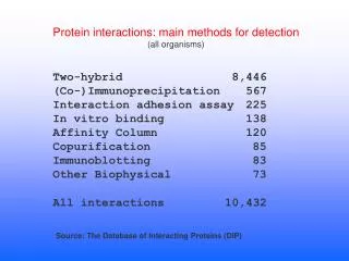
Protein-protein interactions • Two hybrid system • Co-immunoprecipitation • Surface plasmon resonance • Protein arrays • Protein crosslinking • FRET- fluorescence Resonance Energy Transfer
Immunoprecipitation • Specific Ab binds protein in solution • Solution is eluted over Protein A column • Protein is eluted from Ab • Co-IP • Also allows for study of proteins bound to IP’d protein • ID protein complexes
SPR • Surface plasmon resonance • Biomolecular interaction analysis • BIACORE • Protein is immobilized onto surface • Light is refracted onto thin metal layers • Immobile protein refractive index changes when ligand is bound
Protein arrays • ELISA based format: • Ab, proteins, peptides immobilized • Solution to be searched is layered on top • Binding of partner proteins is detected by SPR or fluorescence
Protein crosslinking • X-linking agent “locks” interacting proteins • Formaldehyde • Linking is highly specific • Can be performed in vivo • Cell extract can be subjected to IP assays • Identify x-linked proteins via co-IP • Chromatin IP – ID bound DNA sequences
Fluorescent protein tags • Protein-protein interactions with fluorescence energy transfer (FRET) • Visualizing protein localization • Green fluorescent protein (GFP)