Download
1 / 33
330 likes | 341 Vues
Protein Methods. Andy Howard Introductory Biochemistry, Fall 2014 17 September 2014. Protein methods and functions. Today we ’ l l finish our discussion about how we learn about proteins. Plans for Today. Purification Methods Structure methods Crystallography NMR CryoEM

E N D
Protein Methods Andy HowardIntroductory Biochemistry, Fall 2014 17 September 2014 Protein Methods
Protein methods and functions • Today we’ll finish our discussion about how we learn about proteins Protein Methods
Plans for Today • Purification Methods • Structure methods • Crystallography • NMR • CryoEM • CD, Spectroscopy Protein Methods
Affinity chromatography • Stationary phase contains a species that has specific favorable interaction with the protein we want • DNA-binding protein specific to AGCATGCT: bind AGCATGCT to a column, and the protein we want will stick; every other protein falls through • Often used to purify antibodies by binding the antigen to the column Protein Methods
Metal-ion affinity chromatography • Immobilize a metal ion, e.g. Ni, to the column material • Proteins with affinity to that metal will stick • Wash them off afterward with a ligand with an even higher affinity • We can engineer proteins to contain the affinity tag:poly-histidine at N- or C-terminus Protein Methods
High-performance liquid chromatography • Many LC separations can happen faster and more effectively under high pressure • Works for small molecules • Protein application is routine too, both for analysis and purification • FPLC is a trademark, but it’s used generically Protein Methods
Electrophoresis • Separating analytes by charge by subjecting a mixture to a strong electric field • Gel electrophoresis: field applied to a semisolid matrix • Can be used for charge (directly) or size (indirectly) Protein Methods
SDS-PAGE • Sodium dodecyl sulfate: strong detergent, applied to protein • Charged species binds quantitatively • Denatures protein • Good because initial shape irrelevant • Bad because it’s no longer folded • Larger proteins moveslower because theyget tangled in the matrix • Log(MW) = b - av,where v is the velocity b Log(molwt) b/a Velocity Protein Methods
SDS PAGE illustrated Protein Methods
Isoelectric focusing I • Protein applied to gel without charged denaturant • Electric field set up over a pH gradient (typically pH 2 to 12) • Protein will travel until it reaches the pH where charge =0 (isoelectric point) Protein Methods
Isoelectric focusing II • Sensitive to single changes in charge (e.g. asp asn) • Can be readily used preparatively with samples that are already semi-pure Protein Methods
Ultraviolet spectroscopy • Tyr, trp absorb and fluoresce:abs ~ 280-274 nm; f = 348 (trp), 303nm (tyr) • Reliable enough to use for estimating protein concentration via Beer’s law (A = Cl ) • UV absorption peaks for cofactors in various states are well-understood • More relevant for identification of moieties than for structure determination • Quenching of fluorescence sometimes provides structural information Protein Methods
Mass spectrometry • Enables calculation of mass to charge ratio for molecules or fragments • 1980’s: techniques for aerosolizing proteins were developed so that MS could be applied to proteins • Electrospray • Matrix-assisted laser desorption ionization • Useful analytical tool, alone or along with partial digestion by proteases Protein Methods
Warning: Specialty Content! • I determine protein structures (and develop methods for determining protein structures) as my own research focus • So it’s hard for me to avoid putting a lot of emphasis on this material • But today I’m allowed to do that, because it’s one of the stated topics of the day. Protein Methods
How do we determine structure? • We can distinguish between methods that require little prior knowledge (crystallography, NMR, CryoEM)and methods that answer specific questions (XAFS, fiber, …) • This distinction isn’t entirely clear-cut Protein Methods
Crystallography: overview • Crystals are translationally ordered 3-D arrays of molecules • Conventional solids are usually crystals • Proteins have to be coerced into crystallizing • … but once they’re crystals, they behave like other crystals, mostly Protein Methods
How are protein crystals unusual? • Aqueous interactions required for crystal integrity: they disintegrate if dried • Bigger unit cells (~10-40 nm, not 0.2 - 1nm) • Small # of unit cells and static disorder means they don’t scatter terribly well • So using them to determine 3D structures is feasible but difficult Protein Methods
Crystal structures: Fourier transforms of diffraction results • Experiment: • Grow crystal, expose it to X-ray • Record diffraction spots • Rotate through small angle and repeat ~180 times • Position of spots tells you size, shape of unit cell • Intensity tells you what the contents are • We’re using electromagnetic radiation, which behaves like a wave, exp(2ik•x) • Therefore intensity Ihkl = C*|Fhkl|2 Protein Methods
What are these Fhkl values? • Fhkl is a complex coefficient in the Fourier transform of the electron density in the unit cell:(r) = (1/V) hklFhkl exp(-2ih•r) • Critical point: any single diffraction spot contains information derived from all the atoms in the structure; and any atom contributes to all the diffraction spots Protein Methods
The phase problem Fhkl bhkl • Note that we said Ihkl = C*|Fhkl|2 • That means we can figure out|Fhkl| = √Ihkl / C • We can’tfigure out the direction of F:Fhkl = ahkl + ibhkl = |Fhkl|exp(ihkl) • This direction angle is called a phase angle • Because we can’t get it from Ihkl, we have a problem: it’s the phase problem! ahkl Protein Methods
Solving the phase problem • Small molecules: use relationships among intensities of related spots to calculate phases • Big molecules: • Add heavy atom to protein and measure the changes to the diffraction • Change the wavelength and measure how that affects the diffraction • Start with a model based on a related structure and calculate how to fit that structure into the new unit cell • After you do that, it’s usually straightforward to calculate αhkl ρ(r) Protein Methods
What can we learn? • Electron density map + sequence we can determine the positions of all the non-H atoms in the protein—maybe! • Best resolution possible: Dmin = / 2 • Often the crystal doesn’t diffract that well, so Dmin is larger—1.5Å, 2.5Å, worse • Dmin ~ 2.5Å tells us where backbone and most side-chain atoms are • Dmin ~ 1.2Å: all protein non-H atoms, most solvent, some disordered atoms; some H’s Protein Methods
What does this look like? • Takes some experience to interpret • Automated fitting programs work pretty well with Dmin < 2.1Å ATP binding to a protein of unknown function: S.H.Kim Protein Methods
How’s the field changing? • 1990: all structures done by professionals • Now: many biochemists and molecular biologists are launching their own structure projects as part of broader functional studies • Fearless prediction: by 2020: • crystallographers will be either technicians or methods developers • Most structures will be determined by cell biologists & molecular biologists Protein Methods
Macromolecular NMR • NMR is a mature field • Depends on resonant interaction between EM fields and unpaired nucleons (1H, 15N, 31S) • Raw data yield interatomic distances • Conventional spectra of proteins are too muddy to interpret • Multi-dimensional (2-4D) techniques:initial resonances coupled with additional ones Protein Methods
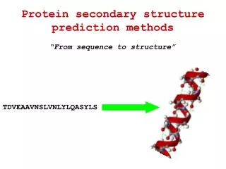
Typical protein 2-D spectrum • Challenge: identify whichH-H distance is responsible for a particular peak • Enormous amount of hypothesis testing required Prof. Mark Searle,University of Nottingham Protein Methods
Results • Often there’s a family of structures that satisfy the NMR data equally well • Can be portrayed as a series of threads tied down at unambiguous assignments • They portray the protein’s structure in solution • The ambiguities partly represent real molecular diversity; but they also represent atoms that area in truth well-defined, but the NMR data don’t provide the unambiguous assignment Protein Methods
Comparing NMR to X-ray • NMR family of structures often reflects real conformational heterogeneity • Nonetheless, it’s hard to visualize what’s happening at the active site at any instant • Hydrogens sometimes well-located in NMR;they’re often the least defined atoms in an X-ray structure • The NMR structure is obtained in solution! • Hard to make NMR work if MW > 55 kDa Protein Methods
What does it mean when NMR and X-ray structures differ? • Lattice forces may have tied down or moved surface amino acids in X-ray structure • NMR may have errors in it • X-ray may have errors in it (measurable) • X-ray structure often closer to true atomic resolution • X-ray structure has built-in reliability checks Protein Methods
Cryoelectron microscopy • Like X-ray crystallography,EM damages the samples • Samples analyzed < 100Ksurvive better • 2-D arrays of molecules • Spatial averaging to improve resolution • Discerning details ~ 4Å resolution • Can be used with crystallography Protein Methods
Circular dichroism • Proteins in solution can rotate polarized light • Amount of rotation varies with • Effect depends on interaction with secondary structure elements, esp. • Presence of characteristic patterns in presence of other stuff enables estimate of helical content Protein Methods
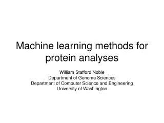
Sperm whale myoglobinPDB 2jho1.4Å16.9 kDa iClicker question 1 • 1. Which protein would yield a more interpretable CD spectrum? • (a) myoglobin • (b) Fab fragment of immunoglobulin G • (c) both would be fully interpretable • (d) CD wouldn’t tell us anything about either protein Anti-fluorescein FabPDB 1flr1.85 Å52 KDa Protein Methods
Ultraviolet spectroscopy • Tyr, trp absorb and fluoresce:abs ~ 280-274 nm; f = 348 (trp), 303nm (tyr) • Reliable enough to use for estimating protein concentration via Beer’s law (A = cl) • UV absorption peaks for cofactors in various states are well-understood • More relevant for identification of moieties than for structure determination • Quenching of fluorescence sometimes provides structural information Protein Methods