Download
1 / 76
980 likes | 1.75k Vues
Bone Marrow Failure/ Aplastic Anemia. Dr. MERVAT A.HESHAM 2008. What is Aplastic Anemia?. Aplastic Anemia is a bone marrow failure disease. Red Blood Cell. Platelets. White Blood Cell. Help to save a Life www.aaaoi.org . Aplastic Anemia patients.

E N D
Bone Marrow Failure/ Aplastic Anemia Dr. MERVAT A.HESHAM 2008
What is Aplastic Anemia? Aplastic Anemia is a bone marrow failure disease. Red Blood Cell Platelets White Blood Cell Help to save a Life www.aaaoi.org
Aplastic Anemia patients • Aplastic Anemia patients have decreased amounts of: - Red Blood Cells - White Blood Cells - Platelets Help to save a Life www.aaaoi.org
Functions of Blood Cells Red Blood Cells Carry oxygen to all body organs White Blood Cells Fight infection and keep you healthy Platelets Help control bleeding Help to save a Life www.aaaoi.org
Symptoms Low Red Blood Cell Fatigue, Headache, Inability to Concentrate Low White Blood Cell Viral Infections, Bacterial Infections Low Platelets Easy Bruising, Nosebleeds, Petichiae Help to save a Life www.aaaoi.org
DEFINITION A disorder of the hemtopoietic system characterized by: Bone marrow - marked reduction of all 3 cell lines Peripheral blood - pancytopenia
PATHOGENESIS Stem cell failure resulting from: 1-An acquired intrinsic stem cell defect 2-An environmental cause Immune mechanisms Growth factor deficiency Defects in the microenvironment
Epidemiology Incidence: 5-10:106 per year Age: 15 –30 years > 60 years Sex: M = F
Hereditary 1-Schwacman – Diamond 2-Fanconi’s anemia syndrome 3-Dyskeratosis congenita Acquired 1-Idiopathic 2- Drugs: dose related idiosyncratic 3-Radiation 4-Chemicals 5-Viruses 6-Pregnancy 7-PNH 8-Disorders of immune system Etiology
Clinical manifestations Insidious onset Manifestations caused by pancytopenia Anemia - weakness, fatigue Thrombocytopenia – bleeding Neutropenia - infections
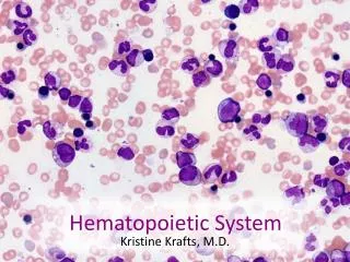
Peripheral blood *Pancytopenia *Normocytic-normochromic anemia *Low reticulocyte index Bone marrow biopsy *Empty fatty spaces *Few hematopoietic cells *Lymphocytes and plasma cells Diagnosis
Bone Marrow Failure Congenital/ Syndromic Acquired
Acquired Aplastic Anemia **Secondary **Idiopathic
Secondary AA 1-Meds/ toxins Chemo Chloramphenicol, benzene, carbamazapine, indomethacin, cimetidine, sulfas, acetazolamide, lithium 2- Radiation 3-Viruses - EBV, HIV, parvo, hepatitis
4-Paroxysmal Nocturnal Hemoglobinurea 5-Malnutrition 6-Myelodysplastic syndromes 7-Thymoma
PATHOPHYSIOLOGY Direct toxic injury to hematopoietic stem cells can be induced by exposure to ionizing radiation, cytotoxic chemotherapy, or benzene. These agents can crosslink DNA and induce DNA strand breaks leading to inhibition of DNA and RNA synthesis.
2-Immune-mediated destruction of hematopoietic stem cells -- Direct killing of the stem cells has been hypothesized to occur via interations between Fas ligand expressed on the T-cells and Fas (CD95) present on the stem cells, which triggers programmed cell death (apoptosis). -- T-lymphocytes also may suppress stem cell proliferation by elaborating soluble factors including interferon-γ.
-T cells from aplastic anemia patients secrete IFN-ã and tumor necrosis factor (TNF). -IFN-ã and TNF are potent inhibitors of both early and late hematopoietic progenitor cells . -Both of these cytokines suppress hematopoiesis by their effects on the mitotic cycle and, more importantly, by the mechanism of cell killing. -Activation of the Fas receptor on the hematopoietic stem cell by the Fas ligand present on the lymphocytes leads to apoptosis of the targeted hematopoietic progenitor cells.
*Cytotoxic T cells also secrete interleukin-2 (IL-2), which causes polyclonal expansion of the T cells. * IFN-ã also induces the production of the toxic gas nitric oxide, diffusion of which causes additional toxic effects on the hematopoietic progenitor cells.
Suppress proliferation with ligand, signals apoptosis Young NEJM 1997
Idiopathic AA *70% or more of cases Higher in SE Asia M = F
AA - Clinical ** Symptoms are due to pancytopenia: pallor, mucosal bleeding, ecchymoses, or petechiae and bacterial or fungal infections.. ** Hepatosplenomegaly and lymphadenopathy do not occur; their presence suggests an underlying leukemia. ** Hyperplastic gingivitis is also a symptom of aplastic anemia.
AA - Labs No RBC = pale, tachycardic No plt = bruising, bleeding No WBC = infection Retic < 1% Plt < 20,000 ANC < 500
AA - Labs Marrow : < 25% cellularity
AA - Evaluation *CBC w/ diff and retic *Bone marrow *Send DEB (Fanconi’s test) *Send Hep A, B, C, D titers HIV *Test for PNH (CD55, CD59) *HLA typing *Fetal hemoglobin *Liver and renal function chemistries
*Quantitative immunoglobulins, C3, C4, and complement. * Autoimmune disease evaluation: Antinuclear antibody (ANA), total hemolytic complement (CH50), Coombs’ test. * HLA typing: Patient and family done at the time of diagnosis of severe aplastic anemia to ensure a timely transplant.
Treatment Options Bone Marrow Transplant GrowthHormones Immune Suppressive Therapy Supportive Care Help to save a Life www.aaaoi.org
TREATMENT 1-Withdrawal of the etiologic agent 2-Supportive treatment Blood and platelet transfusion used with caution- sensitization (filtered) 3-Allogeneic BMT -Preferably from sibling -Curative in 60-90% of patients -Applicable only for a third of patients *Immunosuppression Cyclosporin + ATG Corticosteroids High dose cyclophosphamide *G-CSF/ GM-CSF/ EPO - maybe **Response rate 50-70% Occurs 2-3 months post Rx.
AA Newer *Mycophenolate mofetil (MMF) - cytotoxic to T cells *Monoclonal Ab against IL-2 receptor which is present on activated lymphocytes
AA - Outcomes Age, Younger is better BMT < 20 yr with a sib… 75% 20 - 40 yr with a sib…60% < 20 yr unrelated BMT… 40% 20 - 40 yr unrelated BMT…35% Immunosuppression - 60 - 80% But for how long and consequences…
History: Guido Fanconi Fanconi Anemia (Fanconi pancytopenia syndrome): 1927 - 3 brothers with pancytopenia and physical abnormalities, “perniziosiforme” Fanconi Syndrome (renal Fanconi syndrome): 1936 – Ricketts, growth retardation, proteinuria, glucosuria, and proximal renal tubular acidosis Alter, FA101 (2006)
Fanconi Anemia (FA) Rare (< 1/ 100,000 births) Autosomal recessive Many physical features But up to 20-25% will have no physical findings Mean age at dx 7.8 yrs
Clinical Features Progressive bone marrow failure Most common etiology of inherited bone marrow failure Others include dykeratosis congenita, amegakaryocytic thrombocytopenia, Schwachman-Diamond syndrome Increased risk of MDS and AML (15,000x) Many have monosomy 7, or duplication of 1q (Auerbach et al., Cancer Genet Cytogenet 1991)
Clinical Features Increased risk of solid tumor formation (hepatic, esophageal, oropharyngeal, vulvar) Average age at diagnosis is 23* Cumulative incidence ~30% by age 45** *Shimamura et al., Gene Reviews 2002 (genetests.org) **Alter et al. Blood 2003
FA - genetics Identification of subtypes (compliment groups) A, B, C, D1, D2, E, F, G Identical clinically Sub-units of a common protein/ common pathway Protein modifies FANCD2 FANCD2 interacts with BRCA1 and 2 BRCA1 and 2 needed for DNA repair
PATHOPHYSIOLOGY DNA damage activates a complex consisting of Fanconi proteins A, C, G, and F. This in turn leads to the modification of the FANCD2 protein. This protein interacts, for example, with the breast cancer susceptibility gene BRCA1.
*Fanconi anemia cells are characterized by hypersensitivity to chromosomal breakage as well as hypersensitivity to G2/M cell cycle arrest induced by DNA cross-linking agents. *In addition there is sensitivity to oxygen-free radicals and to ionizing radiation.
Diagnosis -*Pts. with congenital abnormalities are often diagnosed as neonates/infants *Others may be diagnosed when hematological problems occur *Median age of onset of pancytopenia is 7 Usually normal CBC at birth *First develop macrocytosis, then thrombocytopenia, and eventually neutropenia
Diagnosis Based on chromosomal hypersensitivity to cross-linking agents Chromosome fragility test: Mitomycin C (MMC) or diepoxybutane (DEB) added to lymphoctyes – increases the number of chromosome breaks and radial structures Very specific for FA, regardless of severity of disease Can do chromosome breakage analysis on amniotic cells, chorionic villus cells or fetal blood


![Heart Failure [HF]](https://cdn2.slideserve.com/5103874/heart-failure-hf-dt.jpg)