Download
1 / 16
170 likes | 379 Vues
Electrical Activity of Heart. Presentation by: Tarique Ahmad John Frost Grant Post Ian Osborne. Introduction Video. Cardiac Conduction System: http://www.youtube.com/watch?v=zdoSreUAthA. Conducting System. also known as cardiac conducting system or the nodal system

E N D
Electrical Activity of Heart Presentation by: Tarique Ahmad John Frost Grant Post Ian Osborne
Introduction Video Cardiac Conduction System: http://www.youtube.com/watch?v=zdoSreUAthA
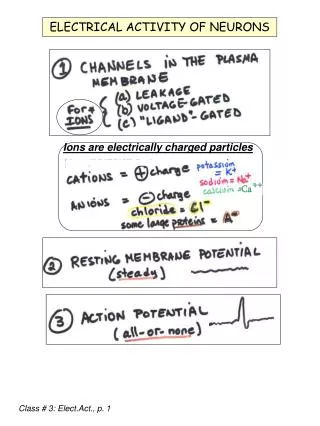
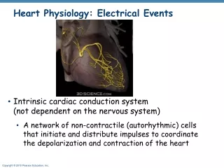
Conducting System • also known as cardiac conducting system or the nodal system • its a network of specialized cardiac muscle cells that initiates and distributes electrical impulses • includes: • Sinoatrial (SA) Node • Atrioventricular (AV) Node • Conducting Cells • Bundle of his (hiss )/ Atrioventricular (AV) Bundle • Purkinje Fibers
Continued... • conducting system allows heart to be automaticity, or autorhythmicity • it contracts on its own, in absence of neural and hormonal stimulation • actual contraction lags behind the electrical pulse • due to time is takes calcium ions to enter the sarcoplasm
http://www.ceufast.com/courses/239/04_Cardiac_Conduction_System.jpghttp://www.ceufast.com/courses/239/04_Cardiac_Conduction_System.jpg
Sinoatrial (SA) Node • In the upper part of the right atrium • specialized bundle of neurons known as the sinoatrial node (SA node) • Acting as the heart's natural pacemaker, • "fires" at regular intervals to cause the heart to beat at a rhythm of 60 to 70 beats per minute
AV / Atrioventricular Node • Specialized Cardiocytes relay the contractile stimulus to the AV bundle, the bundle branches, the Purkinje fibers, and the ventricular myocardium • Located between the atria and the ventricles • There's a 100 millisecond delay once signal is received at AV node
= 100 millisecond long delay Lasts a total of 225 Milliseconds http://washingtonhra.com/16.html
Conducting Cells: Bundle of His • Also known as the AV bundle • Carry the contracting stimulus from the AV node to the Purkinje Fibers • Separates into left and right bundle branches, which are spread across the inner surfaces of the left and right http://www.ambulancetechnicianstudy.co.uk/card.html#.UXHG16KsiSo
Conducting Cells: Purkinje Fibers • Located in the inner ventricle walls of the heart just below the epicardium • Assist the conduction system in the synchronization of contractions • Carry the electrical impulses from the Sinoatrial Node to the Myocardium • Conduct action potentials
Electrocardiogram (ECG/EKG) • graphic record of heart, monitored by electrical activity of heart at certain locations • Electrical events of heart are powerful enough to be detected by electrodes on body surface http://smartmedicalindo.com/product_images/w/452/Schiller_Cardiovit_AT-10_Plus_EKG_Machine__22732_zoom.jpg
Continued... • comparing information from different locations to monitor and check performance of heart • specific components can be checked as well • tracing varies on placement of electrodes, also called leads • used to detect cardiac arrhythmias • abnormal cardiac activity
Understanding an ECG/EKG • P Wave- small wave that accompanies depolarization of atria • QRS Complex- wave that appears after contraction of ventricles • ventricles begin contraction at peak of R wave • T Wave- small wave that indicates ventricular repolarization http://www.usfca.edu/fac-staff/ritter/Image20.gif
Analyzing an EKG • involves measuring size of voltage changes • determining temporal relations between components • focus on amount depolarization during P wave and QRS complex
POP QUIZ!!! • What is the pacemaker of the heart? • Where is it located? • What does AV stand for? • Where is the node located? • What does the bundle of hiss do? • Where are the Purkinje Fibers located? • What does ECG/EKG stand for?
Works Cited Martini, Frederic H., and Edwin F. Bartholomew. Essentials of Anatomy and Physiology. 4th ed. San Francisco: Pearson, 2007. Print. Martini, Frederic H., and Judi L. Nath. Fundamentals of Anatomy and Physiology. 8th ed. San Francisco: Prentice Hall, 2009. Print.