Download
1 / 40
540 likes | 1.11k Vues
Electrical Activity of The HEART . Conduction System of the Heart Dr. Amel Eassawi. Objectives. Students should be able to: Identify the components of the conducting system of the heart. Know the sequence of conduction of impulse in the heart.

E N D
Electrical Activity of The HEART Conduction System of the Heart Dr. AmelEassawi
Objectives Students should be able to: • Identify the components of the conducting system of the heart. • Know the sequence of conduction of impulse in the heart. • Recognize the concept associated with the heart pacemaker. • Appreciate the role of the autonomic nervous system in controlling the rate of generation and conduction of impulse. • Recognize the difference between action potential of SA node and the action potential of ventricular muscle fiber.
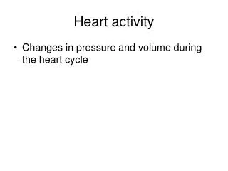
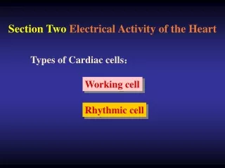
Electrical Activity of Heart Two specialized types of cardiac muscle cells: • Contractile cells • 99% of cardiac muscle cells. • Do mechanical work of pumping. • Normally do not initiate its own action potentials. • Autorhythmic cells • Do not contract. • Specialized for initiating and conducting action potentials responsible for contraction of working cells.
AUTORHYTHMICITY • Heart beats rhythmically as a result of action potentials it generates by itself (Autorhythmicity). • Cardiac autorhythmic cells do not have resting potential instead they show PACE MAKER POTENTIAL • Membrane potential slowly depolarizes between action potential until threshold is reached. • This spontaneous depolarization to threshold is known as PACE MAKER POTENTIAL
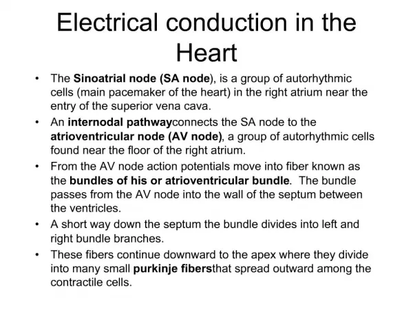
Cardiac Autorhythmic cells • Action potential spread through the myocardial cells through gap junctions. • Impulses cannot spread to ventricles directly because of fibrous tissue. Conduction Pathway: • SA node. • AV node. • Bundle of His. • Purkinje fibers.
Cardiac Autorhythmic cells Cause of Prepotential • Na+ going inside • Ca++ going inside • ↓ K+ going outside • After Prepotential, Depolarization and Repolarization Cause of Depolarization - Ca++ going inside Cause of Repolarization - K+ going outside
Specialized Conduction System of Heart • Sinoatrial node (SA node) • Small mass of specialized cells located in right atrial wall near opening of superior vena cava. • Pacemaker of the heart • Atrioventricular node (AV node) • Small bundle of specialized cardiac cells located at base of right atrium near septum • Bundle of His (atrioventricular bundle) • Cells originate at AV node and enters interventricular septum • Divides to form right and left bundle branches which travel down septum, curve around tip of ventricular chambers, travel back toward atria along outer walls • Purkinje fibers • Small, terminal fibers that extend from bundle of His and spread throughout ventricular myocardium
Specialized Conduction System of Heart SA Node is the Pacemaker of the Heart • SA node discharge rate is high 70-80/min. • This 70-80 action potential/min drive the rest of the heart, therefore, it is known as pacemaker of the heart.
Specialized Conduction System of Heart • Other autorhythmic tissue are firing at slow rate. • They can work as pacemaker, if SA-Node is not functioning e.g. if AV Node takes over as pace-maker, heart rate will be about 50/min. • Any pace-maker other than SA-Node is called ‘Ectopic Pace-maker’. ( associated with organic heart disease or lack of sleep, anxiety, excess caffeine, nicotine)
SPREAD OF CARDIAC EXCITATION Sinoatrial node AV node Bundle of His Bundle Branches Purkinje fibers
SPREAD OF CARDIAC EXCITATION • Cardiac impulse originates at SA node and spread to the atria through the gap junction. Atrial Syncytium, therefore, both atria depolarize at the same time. • Impulse (action potential) travel to AV node by Internodal pathway. • AV node is the only point of electrical contact between atria and ventricle (as atria and ventricle are separated by fibrous ring which is non-conductive).
SPREAD OF CARDIAC EXCITATION AV Node delay • At AV node, there is delay of 0.1 sec (100 milli- sec). This delay is important to allow complete ventricular filling. • because it allows the atria to contract and empty their blood into the ventricle, before impulse reaches the ventricle and causes ventricular depolarization and contraction.
SPREAD OF CARDIAC EXCITATION Ventricular Excitation • After AV node delay of 0.1sec, impulse (AP) travels quickly via Right Bundle Branch and Left Bundle Branch [branches of Bundle of His] to Purkinje Fibers to the ventricles. • Both ventricle depolarize, than contract at same time. • Conduction in Purkinje Fiber is fastest 2-4 meter/sec, therefore, both ventricle depolarize quickly and at the same time. Why Conduction is slow at AV-Node? • Because there are less gap junctions. • Diameter of the fiber is small.
Conduction speed in cardiac tissue Slowest Conduction at AV node Fastest Conduction at purkinje fibers
Abnormal conduction Sino-atrial block: The sinus node fires but the stimulus does not excite the atria because it is blocked at the junction between the two. AV junctional block: The blocks at the level of the AV junction are classified according to the severity of the block. First-degree block: all action potentials are conducted through the AV junction but the velocity of conduction is depressed.
Abnormal conduction Second-degree block: some of the action potentials are not conducted at all through the junction and therefore do not excite the ventricles. Third-degree block: all action potentials are completely blocked at the AV junction and the ventricles are not stimulated by the atria. This would lead to cardiac arrest unless a latent pacemaker escapes and drives the ventricles.
Abnormal conduction Bundle Branch block: Conduction at one of the branches become blocked. The wave of excitation spread from the intact branch to depolarize the whole ventricle, which take longer time than if both branches are intact. • Right bundle branch block. • Left bundle branch block.
Abnormal conduction APPLIED: If no conduction occurs from SA Node to the ventricle through AV node, therefore, atrial rate is separate [75/min] from the ventricular rate which follows the Purkinje fibers and is about 30/min. • IMPORTANT If ventricular rate is very slow e.g. complete heart block, we need artificial pacemaker [implanted device which generates impulse].
Myocardial Action Potential • Once myocardial cells are stimulated by action potential originating in SA node, it produces its own action potential. • Ventricular Muscle membrane has resting membrane potential of -90mV. • Action Potential of ventricular muscle fiber has four phases 0, 1, 2, 3 ,4.
Myocardial Action Potential Ventricular Action Potential • Rapid depolarization (Phase 0) – due to Na+ influx • Rapid Repolarization (Phase 1) - Due to closure of Na+ channels • Slow depolarization (Phase 2) - this is called Plateau phase and is maintained for 200 – 300 ms – due to Ca++ influx • Repolarization (Phase 3) – due to K+ efflux • Resting Membrane Potential (Phase 4)
Phase 0 = due to Ca++ influx not Na+ Phase 1= absent Phase 2= Less prominent
Action Potential Configurations from Different Regions of the Mammalian Heart Pacemaker Cells Vs. Contractile Cells Fast Fibers vs. Slow Fibers Action potential
Relationship of an Action Potential and the Refractory Period to the Duration of the Contractile Response in Cardiac Muscle
refractory period • Because long refractory period occurs in conjunction with prolonged plateau phase, summation and tetanus of cardiac muscle is impossible • Ensures alternate periods of contraction and relaxation which are essential for pumping blood
Effect of Sympathetic and parasympathetic Stimulation on Pacemaker Potential • Epinephrine, norepinephrine, adrenaline and noradrenaline causes prepotential to occur faster therefore increase the heart rate. • Acetylcholine causes prepotential to occur at slow rate therefore decrease the heart rate.
Effect 0f Sympathetic Stimulation on Pacemaker Potential Why Sympathetic Stimulation causes Prepotential to occur faster? • Because Sympathetic Stimulation causes - more Na+ influx [entry] - more Ca2+ influx [entry] - decreased K+ efflux [going outside] • Therefore, membrane potential changes quickly from -60mV to -40mV [increases the slope of Prepotential] and when it reaches the threshold level, AP starts.
Effect 0f parasympathetic Stimulation Pacemaker Potential Why parasympathetic causes Prepotential to occur after long time? • Because Parasympathetic Stimulation causes - decreased Na+ influx [entry] - decreased Ca2+ influx [entry] - increased K+ efflux [going outside] • Therefore, membrane potential changes slowly from -60mV to -40mV [decreases the slope of Prepotential] and when it reaches the threshold level, AP starts.
Control of heart rate • Heart rate is determined by balance between Inhibition of SA node by the parasympathetic (vagus nerve)and stimulation by sympathetic Under resting condition parasympathetic discharge dominates POINT TO PONDER In Transplanted Heart, where there is no sympathetic and parasympathetic nerve supply, what will be the rate of SA Node discharge [Heart Rate] ?
References • Human physiology, Lauralee Sherwood, seventh edition. • Text book physiology by Guyton &Hall,11th edition. • Physiology by Berne and Levy, sixth edition.