Download

1 / 38
520 likes | 1.1k Vues
Applications of Molecular Cytogenetics. Dr Mohammed Alqahtani CSLT(CG), CLSp(CG), RT,MBA, Ph.D Genomic Medicine Unit Founder & Director Center of Excellence in Genomic Medicine Research Founder & Director . Lecture Objectives.

E N D
Applications of MolecularCytogenetics Dr Mohammed Alqahtani CSLT(CG), CLSp(CG), RT,MBA, Ph.D Genomic Medicine Unit Founder & Director Center of Excellence in Genomic Medicine Research Founder & Director
Lecture Objectives • Understand how molecular cytogenetic techniques can be used to identify clinically relevant chromosome abnormalities • Be aware of the different types of molecular techniques that can be used to identify and clarify chromosome rearrangements
Molecular Cytogenetic Techniques Powerful complement to conventional cytogenetic analysis of: • aneuploidy • structural rearrangements • submicroscopic rearrangements • microdeletions/duplications • subtelomere rearrangements
Patient Basic chromosomal analysis Family of the patient Molecular cytogenetic analysis Molecular biological analysis
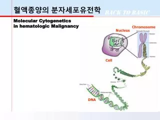
hybridization PCR Molecular cytogenetic examinations • In most of cases interphase cells could be used for analysis (with exception of whole chromosome painting probes and M-FISH) • Examples of methods: • in situ hybridization and its modifications (CGH, M-FISH, fiber FISH atd.) • Gene chips, resp. array CGH, DNA microarray etc. • PRINS, PCR in situ • quantitative fluorescent PCR, real time PCR • methods based on amplification of probe attached to target sequence (MLPA, MAPH)
Molecular Cytogenetics Era • 1988 FISH • 1992 Comparative Genomic Hybridization • 1994 Reverse FISH • 1996 Spectral Karyotyping, M-FISH • 1999 M-Band analysis • 2002 Fiber FISH • 2002 Primed in situ labeling (PRINS) • 2002 Microarray
Molecular Cytogenetic testing • POSTNATAL Stat Blood Routine Blood Skin Biopsy Product of Conception
PRENATAL Amniotic Fluid Chorionic Villus Sampling Fetal Cord Blood • CANCER GENETICS Bone Marrow Oncology Blood Solid Tumor Lymph Node Pleural Effusion Core Biopsy
Molecular Application • FISH • CGH • PCR • Real Time PCR • DNA Sequencing • Microarray
FISH • A technique that hybridizes a DNA nucleic acid probe to a target DNA sequence contained within a cell nucleus. • A variety of specimen types can by analyzed using FISH. The intact cells are attached to a microscope slide using standard cytogenetic methods.
(FISH) TO RULE OUT: • Chromosome Microdeletion Detection • Interphase Chromosome Enumeration • Gene Rearrangements (ie, bcr/abl, PML/RARA) • Cryptic Chromosomal Rearrangements • Marker Chromosome Identification • Chromosome Breakpoint Mapping
FISH for Detection of Single to Multiple Genetic Events Dual Targets Two colors Multiple Targets Multi- colors Single Target One color Allows one to look at multiple genomic changes within a single cell, without destruction of the cellular morphology.
Probes • Probe is a nucleic acid that • can be labeled with a marker which allows identification and quantitation • will hybridize to another nucleic acid on the basis of base complementarity
Probes Types of labeling • Direct & Indirect • Radioactive (32P, 35S, 14C, 3H) • Fluorescent • FISH: fluorescent in situ hybridization • Biotinylated (avidin-streptavidin)
Probe • A part of DNA (or RNA) that is complementary to certain sequence on target DNA (i.e. DNA of the patient) • Plasmid, phage DNA, cosmid (or combination of phage and plasmid DNA), YAC • PCR-product (amplification of certain segment of chromosomal DNA)
DIRECT FLUORESCENT -LABELED PROBE F Specimen DNA T A A T C G T A G COVALENT BOND A G C T C F FISH Probe DNA
Types of FISH Probes • Centromere • Telomere • Whole chromosome paint • locus
Types of probes Centromeric (satellite) probes Locus specific probes Whole chromosome painting probes
Types of probes • Telomeric probes have specificity for a single human chromosome arm. They contain a locus estimated to be within 300 kb of the end of the chromosome. • WCP Chromosome Painting Probesthehybridized probe fluoresces with bright intensity along the length of chromosome • CEP Chromosome Enumerator Probes (centromere area) • Most are Alpha and Satellite III Probes • Centromere regions stained brighter - means they are rich in A-T bonds
Types of probes • LSI Locus Specific Identifiers • Deletion Probes • Translocation Probes • Gene Detection & Localization • Gene Amplification Probes
In which conditions we have to indicate FISH analysis? • The material doesn't contain metaphase chromosomes • Unsuccessful cultivation • It isn't possible to cultivate the tissue from patient (preimplantation analysis, rapid prenatal examinations, examinations of solid tumors or autopsy material) • Analysis of complicated chromosomal rearrangements
In which conditions we have to indicate FISH analysis? • Identification of marker chromosomes • Analysis of low-frequency mosaic • Diagnosis of submicroscopic (cryptic) chromosomal rearrangements • Microdeletion syndromes • Amplification of oncogenes and microdeletion of tumor-suppressor genes in malignancies
Multi Color FISH • Multicolor FISH can provide “colorized” information relative to chromosome rearrangements, especially useful in specimens where chromosome preparations are less than optimal for standard cytogenetic banding analysis.
FISH Procedure • Denature the chromosomes • Denature the probe • Hybridization • Fluorescence staining • Examine slides or store in the dark
Hybridization target DNA denaturation hybridization probe
Hybridization • Nucleic acid hybridization is the formation of a duplex between two complementary sequences • Intermolecular hybridization: between two polynucleotide chains which have complementary bases • DNA-DNA • DNA-RNA • RNA-RNA • Annealing is another term used to describe the hybridization of two complementary molecules
Automated Hybridization HYBrite™ • The probe and target DNA are denatured together. • Faster, easier, and safer hybridization.
Visualization of the Probe • DNA probe is labeled with a colored fluorescent molecule. • This fluorescent molecule remains attached to the DNA during the hybridization process • The molecule emits a particular color when viewed through a fluorescence microscope that is equipped with the appropriate filter sets.
CCD Camera Fluorescent Microscope FISH Analysis Software Filters
13 (green) 21 (red) 99.9% correlation X (green), Y (red) 18 (aqua) Results: 24 hours Results: 7 - 10 days FISH vs. Karyotyping