Download

1 / 31
310 likes | 449 Vues
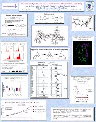
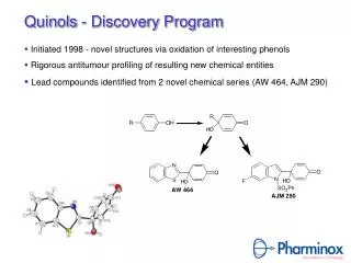
Novel Antitumour Quinols Charles Matthews. Introduction: Heteroaromatic Quinols. Discovered by Malcolm Stevens’ CRUK group at University of Nottingham through oxidation of bioactive phenols Quinols contain a 4-hydroxycyclohexa-2,5-dienone moiety

E N D
Novel Antitumour Quinols Charles Matthews
Introduction: Heteroaromatic Quinols • Discovered by Malcolm Stevens’ CRUK group at University of Nottingham through oxidation of bioactive phenols • Quinols contain a 4-hydroxycyclohexa-2,5-dienone moiety • Michael Acceptors: covalently bind with nucleophiles e.g. thiols
Introduction: PMX 464 • Initially synthesised by Dr Geoff Wells • NCI 60 Cell Line Screening • Potent: nanomolar GI50s • Novel pattern of activity with selectivity for Colon, Breast and Renal lines at LC50 level • COmputer PAttern REcognition (COMPARE) negative with all known chemotherapeutic agents • COMPARE positive with other inhibitors of thioredoxin/thioredoxin reductase (Trx/TR) system. • Antitumour activity in renal and colon xenograft models • Orally bioavailable, water soluble
Cancer drug resistance (e.g. cisplatin) Antioxidant (removal of H2O2) Growth stimulation DNA synthesis (H donor for ribonucleotide reductase) Transcription factor regulation (p53, NF-kB, AP-1, Hif1) Inhibition of apoptosis (ASK1 complexation) Introduction: Thioredoxin System and Cancer • Over-expression of Trx1 in half tumours investigated (e.g. colon, liver, lung), correlates with tumour aggression and resistance to therapy • Promotes tumour growth, co-cytokine • Antioxidant, regulates activity of important growth factors • Reduced apoptosis in tumour cells where Trx1 is overexpressed • Trx1 inhibits apoptosis signal-regulating kinase-1 (ASK1) preventing tumour cell apoptosis in response to cellular stressors.
HCT116: 16 h 1 μM PMX 464 Early apoptotic DMSO 1 3 6 PMX 464 (μM) Late apoptotic/necrotic Propidium Iodide (red) annexin V-FITC (green) PARP cleaved PARP caspase 3 cleaved caspase 3 Introduction: PMX 464 Induced Apoptosis • Apoptosis induction confirmed with annexin V-FITC/propidium iodide assay and western blotting for PARP and caspase 3 cleavage • Intrinsic apoptotic pathway activated
Introduction: PMX 464 Mechanism of Action • PMX 464 inhibits the Trx1/TR-dependent reduction of insulin with an IC50 of 28 M • IC50 reduced to 2.7 M with pre-incubation of Trx/TR with PMX 464 prior to addition of insulin suggests irreversible inhibition • 3, 4 or 5 molecules of PMX 464 shown bound to human Trx1 by mass spectrometry • Trypsin digest of the PMX 464 bound Trx1 revealed either one or two molecules of PMX 464 bound to fragment containing the active site cysteines of thioredoxin. • TR gene and protein levels upregulated following treatment of HCT116 cells. • Bradshaw, T.D., et al., Elucidation of thioredoxin as a molecular target for antitumor quinols. Cancer Res, 2005. 65(9): p. 3911-9. -Cys-Gly-Pro-Cys- Conserved active site of Trxs
Introduction: Effects of PMX 464 on Cellular Redox Homeostasis • A transient increase in cellular levels of ROS is apparent 3-7 h after treatment with PMX 464 in the most sensitive cell line HCT116 mM PMX464 • Intracellular glutathione (GSH) levels increase in response to quinols • Depletion of GSH increased the sensitivity of cells whilst supplementation with a GSH precursor reduced quinol activity • Thus GSH may serve to protect the cells from quinol induced oxidation of protein thiols
Threonine 838 N-terminal coiled coil domain C-terminal coiled coil domain S—S Trx1 ASK1 ASK1 N C N C SH—SH Trx1 T838 T838 Trx1 SH—SH T838 ASK1 T838 ASK1 N C C N Inactive Trx1 S—S H2O2, TNF-, cisplatin TRAF2/6 TRAF2/6 TRAF2/6 TRAF2/6 ASK1 ASK1 C C N N T838 T838 TRAF2/6 TRAF2/6 TRAF2/6 TRAF2/6 P P TRAF2/6 TRAF2/6 TRAF2/6 TRAF2/6 T838 T838 ASK1 ASK1 N C N C Active Introduction: Apoptosis Signalling Kinase (ASK1) • ASK1 signalling is involved in apoptosis induction in response to TNF-α and H2O2 • The ASK1 signalosome is inactive when Trx1 is bound. • Cellular stressors cause oxidation of Trx1. • Trx1 dissociates and TRAF2/6 are recruited. • ASK1 activated by trans-auto phosphorylation. • ASK1 phosphorylates MEKKs which phosphorylate JNK and p38 MAPKs but not ERK1/2. • Sustained (>30min) JNK activation shown to be pro-apoptotic
Hypothesis: Quinols cause dissociation of thioredoxin from ASK1 leading to the induction of apoptosis through activation of downstream MAP Kinases.
Whole Cell Lysates IP: Trx1 ASK1 Trx1 Whole Cell Lysates IP: V5 EV Trx1-V5 EV Trx1-V5 DMSO DMSO 5 M 5 M DMSO DMSO 5 M 5 M -Ab +Ab PMX PMX -Ab +Ab PMX PMX 290 464 290 464 ASK1 (lower band) V5 DMSO DMSO 5 M 5 M DMSO DMSO 5 M 5 M PMX PMX PMX PMX 290 464 290 464 Results: Dissociation of ASK1 From Trx1 • Immunoprecipitation using anti-Trx1 monoclonal antibody and protein A/G agarose beads, HCT116 cells. • ASK1 coimmunoprecipitated with Trx1 • PMX 464 and 290 cause dissociation of ASK1 from Trx1 • Similar results in MCF7 (breast) and A549 (lung) cell lines • No antibody in the negative control sample • ASK1 could have been bound to antibody non-specifically • HCT116 cells transfected with epitope-tagged (V5) Trx1 • Immunoprecipitation repeated with anti-V5 antibody
A IP: Trx1 IB: ASK1 IP: Trx1 IB: Trx1 0 5 60 180 360 min 5mM PMX 464 DMSO 0.5 1 5 10 PMX 464 (mM) Results: Time/Dose Dependence of ASK1 Dissociation From Trx1 • HCT116 cells treated with increasing doses of PMX 464 for 3h or with 5 mM PMX 464 for varying time points • Time and dose dependence of ASK1 dissociation from Trx1 demonstrated
HCT116 MCF7 phospho JNK total JNK actin DMS0 0.5 1.0 3.0 5.0 10.0 PMX 464 (M) 0 5 30 60 180 360 min 5 M PMX 464 DMS0 0.5 1.0 3.0 5.0 10.0 PMX 464 (M) 0 5 30 60 180 360 min 5 M PMX 464 phospho p38 total p38 actin 0 5 30 60 180 360 min 5 M PMX 464 DMS0 0.5 1.0 3.0 5.0 10.0 PMX 464 (M) 0 5 30 60 180 360 min 5 M PMX 464 DMS0 0.5 1.0 3.0 5.0 10.0 PMX 464 (M) Results: Activation of MAP Kinase Pathways Downstream of ASK1 • Phospho-specific antibodies used to determine MAP kinase activation. • Both JNK and p38, the MAP kinases downstream of ASK1, were activated in HCT116 and MCF7 cells following treatment with apoptosis inducing doses of PMX 464 • Activation occurred as early as 5 min post-treatment, peaking between 1 and 3 h but is reduced 6 h post-treatment. This is similar for both MAPKs in both cell lines.
HCT116 phospho ERK total ERK DMS0 0.5 1.0 3.0 5.0 10.0 PMX 464 (M) 0 5 30 60 180 360 min 5 M PMX 464 Results: ERK activation • ASK1 activation tick list • JNK activation • p38 activation • NO ERK activation • Unexpected ERK activation seen in both HCT116 and MCF7 cells • Different kinetics? • Stronger early onset of activation and more rapid inactivation
Early Apoptosis * Total Cell Death * Results: Pharmacological MAPK inhibitors on Quinol induced apoptosis • Annexin V-FITC/propidium iodide assay • HCT116 treated for 24h with 2 mM PMX 464 ± 1 h pre-incubation with 10 mM MAPK inhibitor • No significant effects on early apoptosis induction or total cell death • * signifies p <0.05 (ANOVA)
- - - - - - - - - - + + - - - - - - + + + + + + - - - - - - - - + + + + - - - - - - - - + + + + Results: Pharmacological MAPK Inhibitors Phospho ERK Total ERK DMSOPMX 464 UO126 10 mM UO126 25 mM • MEK inhibitors do prevent ERK activation • Currently no similar experiment for JNK or p38 inhibitors Phospho ERK Total ERK DMSOPMX 464 PD98059 10 mM PD98059 25 mM
Results: Dominant Negative ASK1 and Trx1 siRNA • Lysine 709 to arginine mutation yields a catalytically inactive, dominant negative ASK protein (ASK K709R) • ASK1 transfection does not activate JNK in HCT116 • Increased ASK1 phosphorylation is seen in response to H2O2 but not PMX 464. Other redox sensitive inhibiors! • H2O2 or PMX 464 induced JNK activation is not prevented by ASK K709R over-expression • MTT Assay to assess GI50s • No effect on PMX 464 activity when Trx1 is knocked down • No effect on PMX 464 activity when ASK K709R is overexpressed DF1 Neg Trx1 Trx1 1 2
Hypothesis: Quinols cause dissociation of thioredoxin from ASK1 leading to the induction of apoptosis through activation of downstream MAP kinases. • Conclusions? • Quinols do cause dissociation of thioredoxin from ASK1 but this does not lead to its autophosphorylation and activation • Downstream MAP kinases are activated but independently of ASK. • Dominant negative protein failed to prevent their phosphorylation • ERK was activated which is not downstream of ASK1 • Apoptosis appears to be independent of this MAP kinase activity • MAPK inhibitors do not significantly affect PMX 464 induced apoptosis
← cytoplasmic catalase (CAT) Con EV1 EV2 1 2 3 4 5 6 7 8 ← V5 mitochondrial catalase (mCAT) Results: Importance of ROS Induction • Transient increase in ROS seen in HCT116 cells with H2DCFDA • Catalase is an enzyme found in cells that can “mop up” excess H2O2 • Lentiviral expression system used to develop HCT116 cell lines that stably overexpress cytoplasmic or mitochondrial catalase • HCT116 EV1 (empty vector), CAT5 (cytoplasmic catalase), mCAT4 (mitochondrial catalase) cell lines developed. Immunofluorescence image of HCT116 mCAT4 showing co-localisation of V5-tagged catalase and Mitotracker Red. Nuclei are counterstained with DAPI.
Results: HCT116 Catalase and quinol activity • MTT assay shows catalase overexpression has no effects on quinol activity but does significantly reduce H2O2 effects * * * = p < 0.05, Student’s t-test
* * Results: HCT116 Catalase and quinol activity • Oxidation of cell-permeable H2DCFDA to fluorescent DCF used to analyse intracellular ROS by flow cytometry. • H2O2-induced intracellular ROS levels were significantly reduced in catalase over-expressing cells. • Quinol-induced intracellular ROS levels were unaffected by over-expression of catalase. PMX 464 H2O2 * = p < 0.01, Student’s t-test • PMX 464 does not directly oxidise H2DCFDA to DCF.
Discussion • Catalse overexpression has little effect on quinol activity • Catalase overepression does not reduce H2DCFDA detectable ROS caused by PMX 464 • Other ROS? • H2DCFDA also detects peroxyl radicals and peroxynitrite anions
PMX 464 is dropped! • PMX 464 shows tumour growth promotionin vivo in an MDA435 xenograft model • It’s thought that PMX 464 is actually metabolised by Trx1 to a phenolic benzothiazole (Gareth Hall) that has been previously shown to have mitogenic and oestrogenic properties • PMX290 now the lead compound
Introduction: PMX 290 • Second generation quinols discovered rather serendipitously in an effort to make an indole series of quinols. • Arylsulfonyl protecting group was tricky to remove, was tested none-the-less. • Indole series more potent than PMX 464, PMX 290 being the most potent. • PMX 290 synthesised by Andrew McCarroll • Broader range of activity including melanoma cell lines • Less water soluble with poorer biovailability in vivo than PMX 464 • Pro-drugs under development at Pharminox to improve pharmaceutical properties • Recently shown to inhibit TrxR > Trx1 cell lysates through interaction with selenocysteine residue of TrxR (Eng-Hui Chew, in press)
Introduction: PMX 290 • Endoplasmic Reticulum (ER) Stress inducer? • Microarray results 500nM PMX 290 on HCT116 • Upregulation of genes involved in ER stress response (pink bars), which itself can increase in intracellular ROS • Several markers of oxidative stress e.g. haem oxygenase1 • Upregulation of BIRC3 (IAP) that can inhibit TRAF2 binding to ASK (involved in its activation) • Upregulation of DUSP5 dual specificity phosphatase
Introduction: PMX 8079 • Does the compound go to the ER? • Fluorescent compound PMX 8079 • Rapidly gets into cells • Co-localises with ER Tracker Red (Molecular Probes) Brightfield/PMX 8079 • MCF7 100X • 30 min 1 mM ER Tracker red • 1h, 5 mM PMX 8079 Overlay PMX 8079 ER
Dreams of easy target identification • Dreams of easy target identification • Treat cells with fluorescent dansyl quinol • Prepare whole cell lysates • Electrophoresis of whole cell lysates • Look at gel under UV light and see nice bands where quinol is bound • Excise band(s) and use mass spectrometry to identify target protein • Didn’t work as dansyl group is only weakly fluorescent, lysates visibly glow but gels/membranes don’t • Antibodies available to dansyl fluorescent group • Try above experiment but carry out Western blot to see if bands are present where dansyl quinol is bound to protein • If there are bands immunoprecipitate dansyl quinol bound proteins from cells treated with fluorescent quinol • Electrophoresis and Western blot of immunoprecipitates • Scale up so bands can be coomassie stained, excise band(s) and use mass spec to identify target protein(s)
Dansyl western blots and immunoprecipitations (IPs) • whole cell lysate: anti-dansyl immunoblots • dansyl-immunoprecipitations, anti-dansyl immunoblot MCF7 HCT116 MCF7 DMSO PMX DMSO PMX 8079 8079 DMSO PMX DMSO PMX 8079 8079 • Reproducable bands in MCF7 suggesting PMX 8079 is binding to several proteins • No bands in HCT116
MCF7 Trx IP, Dansyl immunoblot M D 8079 8079 290 M D D 290 8079 8079 8079 290 + 290 290 8079 • No 12kDa (Trx1) band apparent (where red rectangle is) suggesting that Trx is not bound by dansyl quinol • Two potential target proteins pulled out with Trx • One where PMX 290 competes, one where it doesn’t. This band may be ASK1. • Lower (ASK1) band not as prominent there in the repeat. • No bands in HCT116 Trx or dansyl IP!! (not shown)
Dansyl quinol questions • Why no bands in HCT116 Western blots or immunoprecipitations? • Metabolism of quinol, dansyl group cleaved in HCT116? • Does this occur in the ER as both cell lines show silmilar staining when treated with dansyl compound? • Is it worth identifying the bands seen in MCF7 cells? • No similar “target” in HCT116 where compound is equipotent • Does dansyl group affect activity? • dansyl version of another quinol showed very similar activity and selectivity to its parent compound (AJM322 and its parent BW114) • Trx not labelled with dansyl quinol • try immunoprecipitation with thioredoxin reductase as EHC has shown that it binds to selenocysteine?
Acknowledgements • Tracey Bradshaw • Thilo Hagen • Eng-Hui Chew • Malcolm Stevens • Geoff Wells • Andrew McCarroll • Everyone in the labs