Download
1 / 18
190 likes | 375 Vues
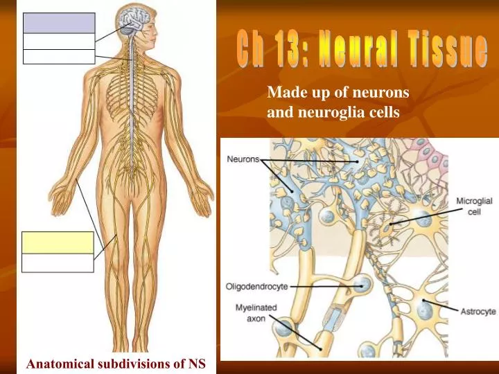
Ch 13: Neural Tissue. Made up of neurons and neuroglia cells. Anatomical subdivisions of NS. CNS Integration, processing and coordination of sensory data and motor commands Higher functions. PNS

E N D
Ch 13: Neural Tissue Made up of neurons and neuroglia cells Anatomical subdivisions of NS
CNS Integration, processing and coordination of sensory data and motor commands Higher functions PNS Sensory or afferent division with sensory neurons. Brings sensory info to CNS. Begins as receptors, ends in? Motor or efferent division with motor neurons.Brings motor commands to peripheral tissue.Ends at effector cells. Functional Overview of NS
Cellular Organization of Neural Tissue Two cell types: • Neurons • Neuroglia • Schwann cells • Satellite cells • Astrocytes • Oligodendrocytes • Microglial cells • Ependymal cells See fig. 13-5 Compare to fig. 14-4
General Neuron Structure • Cell body or Soma with Perikaryon • Dendrites • Axon with axon hillock • Synaptic terminals
Astrocytes: largest & most numerous • Function:BBB • structural framework & repairs • regulation of ions, nutrients, gases
Oligodendrocyte Smaller than astrocyte Produce myelin in CNS (white matter vs. gray matter!) Myelin = ?
Microglia cells • Smallest • Phagocytosis of ? • # during infection or injury
Ependymal cells Lining of ventricles & central canal Some regions ciliated Some specialized to produce CSF
Schwann Cells and Peripheral Axons Responsible for myelination, but surround all peripheral axons! Involved in repair mechanism after injury Wallerian Degeneration myelinated
StructuralNeuron Classification Anaxonic In CNS Unipolar Also called pseudounipolar Sensory neurons Axon hillock See fig. 13-10
Structural Neuron Classification cont. . . Bipolar Unmyelinated Rare, but important in special senses Multipolar Most common All motor neurons
Functional Neuron Classification 1) somatic vs. visceral sensory or afferent monitoring of ? 2) somatic vs. visceral motor or efferentcarry instructions to ? 3) Inter- or association neurons
Synapse Site of communication between two nerve cells or nerve cell and effector cell neuro-effector junctions (example?) Electrical vs. chemical synapses
Space between two cells Signal transduction via NT Most common Direct physical contact between cells = gap junctions Direct signal transduction Rare, but occurs in CNS and heart Chemical Synapsevs.Electrical Synapse
Chem. Synapse Structure • Axon terminal of presynaptic cell • Synaptic cleft • Dendrite or cell body of postsynaptic cell
Neuron Organization • Divergence - One neuron synapses with several, effectively "spreading the word". • Convergence - Several neurons synapse with a single neuron, concentrating the input. • Serial processing - step-wise, sequential • Parallel processing - simultaneous processing of different information
Anatomical Organizatin of NS Collections of cell bodies - ganglion in PNS, center or nucleus in CNS Bundles of axons - tracts in CNS, nerves in PNS “White” = myelinated axons, both nerves and tracts “Gray” = non-myelinated material, dendrites, synapses and cell bodies as well as nonmyelinated axons. In CNS – nucleus; in PNS - ganglia The End Compare to fig. 13-15