Download
1 / 54
580 likes | 812 Vues
Ankle, Lower Leg Lab. BONY PALPATION:. Anterior Aspect . fibular head. fibular shaft. tibial plateau . tibial shaft. Posterior Aspect. medial malleolus. lateral malleolus. calcaneus. SOFT TISSUE PALPATION:. Lateral Aspect. lateral compartment (peroneals).

E N D
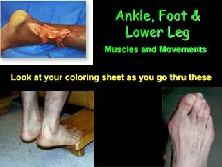
deep posterior muscles • posterior tibialis muscle; flexor digitorum longus muscle; flexor hallucis muscle
anterior tibialis muscle extensor hallucis longus muscle extensor digitorum longus muscle anterior compartment
gastrocnemius muscle superficial posterior compartment
superficial posterior compartment • soleus muscle
Anterior Drawer Test Positioning the Athlete: The athlete is seated on a table with the knee flexed to 90 degrees and the involved foot relaxed in slight plantar flexion. The examiner stabilizes the tibia and fibula with one hand and grasps the calcaneus with the other. Action: While assuring stabilization of the distal tibia and fibula, the examiner applies an anterior force to the calcaneus and talus. Positive Finding: Anterior translation of the talus away from the ankle mortise that is greater on the involved side, as opposed to the noninvolved side, indicates a positive sign for a possible anterior talofibular ligament sprain.
Talar Tilt Test- Inversion Positioning the Athlete: The athlete lies on the uninvolved side on a table with the involved foot relaxed and the knee flexed to approximately 90 degrees. Make sure to stabilize the distal tibia with one hand with grasping the talus with the other. Action: The examiner first places the foot in the neutral plantar flexion and dorsiflexion position (anatomical position). Then tilt the talus into an adducted position. Positive Finding: Range of motion in the adducted position on the involved foot greater than that of the noninvolved foot reveals a positive test. This may be indicative of a tear of the calcaneofibular ligament of the ankle.
Talar Tilt Test- Eversion Positioning the Athlete: The athlete lies on the involved side on a table with the involved foot relaxed and the knee flexed to approximately 90 degrees. Make sure to stabilize the distal tibia with one hand with grasping the talus with the other. Action: The examiner first places the foot in the neutral plantar flexion and dorsiflexion position (anatomical position). Then tilt the talus into an abducted position. Positive Finding: Range of motion in the abducted position on the involved foot greater than that of the noninvolved foot reveals a positive test. This may be indicative of a tear of the deltoid ligament of the ankle.
Kleiger Test-Deltoid Positioning the Athlete: The athlete sits with the leg off of the table and the knee at approximately 90 degrees of flexion. The examiner stabilizes the distal tibia and fibula with one hand and the medial and inferior aspects of the calcaneus with the other. The ankle should be in a neutrally aligned position. Action: The examiner applies an externally rotated force upon the calcaneus. Positive Finding: Complaints of pain along the medial aspect of the ankle when an externally rotated force is applied in neutral dorsiflexion may indicate a deltoid ligament injury.
Kleiger Test- Syndesmosis Positioning the Athlete: The athlete sits with the leg off of the table and the knee at approximately 90 degrees of flexion. The examiner stabilizes the distal tibia and fibula with one hand and the medial and inferior aspects of the calcaneus with the other. The ankle should be in a dorsiflexed position. Action: The examiner applies an externally rotated force upon the calcaneus. Positive Finding: Pain may be present medially and slightly more proximally, indicating distal tibiofibular syndesmotic involvement.
Compression Test Positioning the Athlete: The athlete lies supine with the affected leg extended and the ankle/foot just off the examining table. The examiner stands next to the athlete’s leg and notes where the pain originates. Action: The examiner squeezes the tibia and fibula together at some point away from the painful area. Positive Finding: Pain at the site of injury my be indicative of a fracture. Compression of the two bones may exaggerate pain at the fracture site.