Download
1 / 82
820 likes | 893 Vues
PERIPHERAL & AUTONOMIC NERVOUS SYSTEMS Human Anatomy Sonya Schuh-Huerta, Ph.D. Leonardo Da Vinci, The Battle of Anghiari. The Peripheral Nervous System. The PNS Is the nervous system outside the brain & spinal cord Provides vital links to the body & outside world

E N D
PERIPHERAL & AUTONOMIC NERVOUS SYSTEMS Human Anatomy Sonya Schuh-Huerta, Ph.D. Leonardo Da Vinci, The Battle of Anghiari
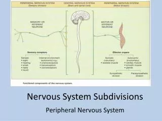
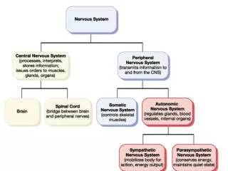
The Peripheral Nervous System • The PNS • Is the nervous system outside the brain & spinal cord • Provides vital links to the body & outside world • Nerves allow the CNS to receive info & initiate action • Sensory inputs & motor outputs • Categorized as • Somatic or visceral • General or special
Central nervous system (CNS) Peripheral nervous system (PNS) Sensory (afferent) division Motor (efferent) division Somatic sensory General: Touch, pain, pressure, vibration, temperature, and proprioception in skin, body wall, and limbs Visceral sensory General: Stretch, pain, temperature, chemical changes, and irritation in viscera; nausea and hunger Somatic nervous system Autonomic nervous system (ANS) Motor innervation of all skeletal muscles Motor innervation of smooth muscle, cardiac muscle, and glands Special: Hearing, equilibrium, vision Special: Taste, smell Sympathetic division Parasympathetic division Functional Organization of the PNS
Basic Structural Components of PNS • Sensory receptors pick up stimuli from • inside or outside body (we’ll cover later) • Nerves & ganglia • Nerves bundles of peripheral axons • Ganglia clusters of peripheral neuronal cell • bodies • Motor endings axon terminals of motor neurons • Innervate effectors (muscle fibers & glands)
The Cranial Nerves – Yes you have to know them… • Attach to brain & pass through foramina of the skull • Numbered I–XII (1-12) • Cranial nerves I & II attach to the forebrain • All others attach to brain stem • Primarily serve head & neck structures • The vagus nerve (X) is the only cranial nerve that extends into abdomen
The Cranial Nerves Filaments of olfactory nerve (I) Frontal lobe Olfactory bulb Olfactory tract Optic nerve (II) Temporal lobe Optic chiasma Optic tract Oculomotor (III) Infundibulum Trochlear (IV) Facial (VII) Vestibulocochlear (VIII) Trigeminal (V) Abducens (VI) Glossopharyngeal (IX) Vagus (X) Cerebellum Accessory (XI) Medulla oblongata Hypoglossal (XII)
The Cranial Nerves Cranial nerves Sensory function Motor function Somatic sensory (SS) Visceral sensory (VS) Somatic motor (SM) Visceral motor: parasympathetic (VM) Odessa Ofelia O’Conner Takes Tests Amazingly! I Olfactory Smell II Optic Vision III Oculomotor SM VM IV Trochlear SM General V Trigeminal SM VI Abducens SM
The Cranial Nerves Cranial nerves Sensory function Motor function Somatic sensory (SS) Visceral sensory (VS) Somatic motor (SM) Visceral motor: parasympathetic (VM) Fiesty Victor Gets Very Agitated Here… VII Facial General General; taste SM VM VIII Vestibulocochlear Hearing; equilibrium Some IX Glossopharyngeal General General; taste SM VM X Vagus General General; taste SM VM XI Accessory SM XII Hypoglossal SM
I ) Olfactory Nerves • Sensory nerves of smell
II) The Optic Nerves • Sensory nerve of vision
III) The Oculomotor Nerves • Innervates 4 extrinsic eye muscles Table 14.2 (3 of 12)
IV) The Trochlear Nerves • Innervates the superior oblique muscle (an extrinsic eye muscle)
The Trigeminal Nerves • Largest of the cranial nerves • Has 3 divisions • Ophthalmic • Maxillary • Mandibular • Cell bodies of sensory neurons located in the trigeminal ganglion • Mandibular division contains motor fibers that innervate the chewing muscles
VI) The Abducens Nerves • Abducts the eyeball innervates lateral rectus muscle
VII) The Facial Nerves • Innervates muscles of facial expression
VIII) The Vestibulocochlear Nerves • Sensory nerve of hearing & balance
IX) The Glossopharyngeal Nerves • Innervates structures of the tongue & pharynx
X) The Vagus Nerves • A mixed sensory & motor nerve • “Wanders” into thorax & abdomen • Parasympathetic innervation of organs
XI) The Accessory Nerves • Unique among cranial nerves • Accessory nerves come from ventral rootlets of spinal cord • Do not arise from the brainstem
XII) The Hypoglossal Nerves • Runs inferior to the tongue • Innervates the tongue muscles
The Spinal Nerves • 31 pairs contain thousands of nerve fibers Formula C8, T12, L5, S5, Cx1 = 31 • Connect to spinal cord • Named for region of vertebral column • 8 pairs cervical nerves (C1–C8) • 12 pairs thoracic nerves (T1–T12) • 5 pairs lumbar nerves (L1–L5) • 5 pairs sacral nerves (S1–S5) • 1 pair coccygeal nerves (Cx1)
Spinal Nerves – Posterior View Cervical plexus Cervical nerves C1 – C8 Brachial plexus Cervical enlargement Thoracic nerves T1 – T12 Intercostal nerves Lumbar enlargement Lumbar nerves L1 – L5 Lumbar plexus Sacral plexus Sacral nerves S1 – S5 Coccygeal nerve Co1 Cauda equina
Spinal Nerves • Branch into dorsal ramus & ventral ramus • Dorsal&ventral rami contain sensory andmotor fibers • Rami communicantes connect to the base of the ventral ramus • Lead to the sympathetic chain ganglia
Spinal Nerves Sensory axon and cell body Dorsal root ganglion Dorsal root Dorsal ramus Nerves Spinal nerve Ventral ramus Ventral root Axon of motor neuron Neuromuscular junction Sensory receptors in skin (e.g., free nerve endings of sensory neuron)
Spinal Nerves Gray matter White matter Ventral root Dorsal & ventral rootlets of spinal nerve Dorsal root Dorsal root ganglion Dorsal ramus of spinal nerve Ventral ramus of spinal nerve Spinal nerve Rami communicantes Sympathetic trunk ganglion
Innervation of the Back • Dorsal rami • Innervate back muscles • Follow a neat, segmented pattern • Innervate a horizontal strip of muscle & skin • In line with emergence point from the vertebral column
Innervation of the Anterior Thoracic & Abdominal Wall • Thoracic region • Ventral rami arranged in simple, segmented pattern • Intercostal nerves supply intercostal muscles, skin, & abdominal wall
Introduction to Nerve Plexuses& Peripheral Nerves • Nerve plexus a network of nerves! • Branch from ventral rami (except T2–T12) • Branch & join with one another • Form nerve plexuses • In cervical, brachial, lumbar, & sacral regions • Primarily serve the limbs • Fibers from ventral rami criss-cross
The Cervical Plexus • Buried deep in the neck • Under the sternocleidomastoid m. • Most are cutaneous nerves • Some innervate muscles of the anterior neck • Phrenic nerve the most important nerve of the cervical plexus • -Innervates diaphragm, mediastinal pleura & • pericardium Control of breathing!
The Cervical Plexus Ventral rami Segmental branches Hypoglossal nerve (XII) Ventral rami: Lesser occipital nerve C1 Greater auricular nerve C2 Transverse cervical nerve C3 Ansa cervicalis C4 Accessory nerve (XI) C5 Phrenic nerve Supraclavicular nerves
Major terminal branches (peripheral nerves) Roots (ventral rami) Cords Divisions Trunks Anterior Musculocutaneous C5 Upper Lateral Posterior Median C6 Medial Anterior Ulnar C7 Middle Posterior Radial C8 Posterior Anterior Lower Axillary T1 Posterior (c) Flowchart summarizing relationships within the brachial plexus The Brachial Plexus & Innervation of the Upper Limb • Brachial plexus lies in the neck & axilla • Formed by ventral rami of C5–C8 • Cords give rise to main nerves of upper limb
Innervation of the Upper Limb • Musculocutaneous • Innervates the biceps brachii & brachialis m. • Median • Innervates anterior forearm muscles & palm • Ulnar • Innervates intrinsic hand muscles & skin of hand
Innervation of the Upper Limb • Radial • Largest branch of the brachial plexus • Innervates muscles of the posterior upper limb • Axillary • Innervates the deltoid & teres minor m.
Major Nerves of the Upper Limb Axillary nerve Humerus Radial nerve Musculocutaneous nerve Ulna Radius Ulnar nerve Median nerve Radial nerve (superficial branch) Dorsal branch of ulnar nerve Superficial branch of ulnar nerve Digital branch of ulnar nerve Muscular branch Anterior divisions Posterior divisions Median nerve Digital branch
Major Nerves of the Upper Limb Axillary nerve Branches of axillary nerve Radial nerve Ulnar nerve (cut) Median nerve (cut) Posterior cutaneous nerve Deep radial nerve Superficial branch of radial nerve Anterior divisions Posterior divisions
The Lumbar Plexus & Innervation of the Lower Limb • Lumbar plexus • Arises from L1– L4 • Main branches innervate the anterior thigh • Femoral nerve innervates anterior thigh muscles • Obturator nerve innervates adductor muscles
The Lumbar Plexus & Nerves Iliohypogastric Ilioinguinal Femoral Lateral femoral cutaneous Obturator Anterior femoral cutaneous Saphenous (c) Distribution of the major nerves from the lumbar plexus to the lower limb
The Sacral Plexus • Arises from spinal nerves L4–S4 • Caudal to the lumbar plexus • Often considered with the lumbar plexus • Lumbosacral plexus
The Sacral Plexus & Innervation of the Lower Limb • Sciatic nerve the largest nerve of the sacral plexus • Actually 2 nerves in one sheath • Tibial nerve innervates most of the posterior lower limb • Common fibular (peroneal) nerve innervates muscles of the anterolateral leg
The Sacral Plexus & Nerves Superior gluteal Inferior gluteal Pudendal Sciatic Posterior femoral cutaneous Common fibular Tibial Sural (cut) Deep fibular Superficial fibular (c) Distribution of the major nerves from the sacral plexus to the lower limb Plantar branches
A Case Study… • Black lab hit in car accident • Initial inability to walk or move limbs • Hospitalized for 1 week • Regained motor control of lower limbs • & front right limb • However, front left limb completely • paralyzed - no sensation or motor control • What spinal nerves, plexus(es) or peripheral • nerves were likely damaged?
Damage to: • Left spinal nerves C5-C8 • Left Brachial plexus • Peripheral nerves: axillary, • musculocutaneous, radial, • median, & ulnar
Innervation of the Skin: Dermatomes • Dermatome an area of skininnervated by cutaneous branches of a single spinal nerve! • Important diagnostic implications • Upper limb • Skin is supplied by nerves of the brachial plexus • Lower limb • Lumbar nerves anterior surface • Sacral nerves posterior surface
Map of Dermatomes C2 C3 C4 C2 C5 C3 C6 C7 C4 C8 T1 C5 T2 T1 T3 T2 T4 T3 T5 T4 T6 T2 T2 T7 T5 T8 T6 T9 T10 T7 C6 C6 T8 T11 C6 C6 T9 T12 C7 C7 L1 T10 S1 C5 L2 C5 C8 C8 L3 T11 S2 L5 L4 S3 T12 S4 L1 L1 C6 C6 S5 S2 C7 C7 C8 S3 C8 L2 L2 S1 S2 S2 S1 L1 L3 L3 L2 L5 L5 L4 L4 L3 L5 L5 L4 S1 S1 L4 L4 L5 L5 S1 (a) Anterior view (b) Posterior view
Disorders of the PNS • Shingles (Herpes zoster) • Viral infection • Inside of neuron cell bodies of peripheral nerves – breaks out & • affects skin of that region - dermatome • Stems from childhood chicken pox • Often brought on by stress • Mostly experienced by those over age 50 Varicella zoster virus (chicken pox) can become dormant in the nerve cell bodies and less frequently in non-neuronal satellite cells of dorsal root, cranial nerve or autonomicganglion, without causing any symptoms. Years or decades after a chickenpox infection, the virus may break out of nerve cell bodies and travel down nerve axons to cause viral infection of the skin in the region of the nerve. The virus may spread from one or more ganglia along nerves of an affected segment and infect the corresponding dermatome causing a painful itching rash. Although the rash usually heals within 2-4 weeks, some sufferers experience residual nerve pain for months or years, a condition called postherpetic neuralgia. Exactly how the virus remains latent in the body, and subsequently re-activates is not understood.
Disorders of the PNS • Migraine headache • Relates to sensory innervation of cerebral arteries • Arteries dilate & compress & irritate sensory nerve endings • Myasthenia gravis • Progressive weakening of skeletal muscles • Autoimmune disorder • Antibodies destroy acetylcholine receptors
Disorders of the PNS • Carpal Tunnel syndrome • Swelling/inflammation & compression of the median nerve of the wrist due to repetitive, non-ergonomic movements • Pain, discomfort, tingling, loss of feeling
The PNS Throughout Life • Spinal nerves form late in week 4 • Each of the 31 pairs of spinal nerves: • Sends motor fibers to an individual myotome • Sends sensory fibers to overlying band of skin • During week 5, nerves reach the organs they innervate
The Autonomic Nervous System (Ch 15)