Download
1 / 68
770 likes | 1.22k Vues
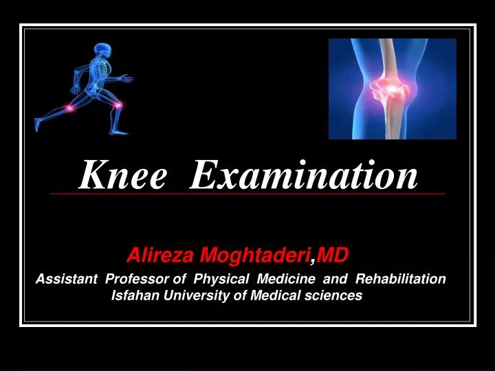
Knee Examination. Alireza Moghtaderi , MD Assistant Professor of Physical Medicine and Rehabilitation Isfahan University of Medical sciences. Knee. The largest joint ,,,,,, Connecting two major bone ,,,,,, More than just a simple Hinge ,,,, Patella is the Largest Sesamoid

E N D
Knee Examination AlirezaMoghtaderi,MD Assistant Professor of Physical Medicine and Rehabilitation Isfahan University of Medical sciences
Knee • The largest joint,,,,,, • Connecting two major bone,,,,,, • More than just a simple Hinge,,,, • Patella is the Largest Sesamoid • Visual examination,,,,, • Unstable joint,,,,, • Patella acts as a Fulcrum,,,,,
Physical exam: Look Feel Move Special tests
Look (always compare) Alignment ( normal, varus or valgus) Effusion Scars Wasting Color
Feel Temperature (compare) Tenderness ( feel the bony prominences) Effusion (fluid collection) * fluctuation * bulging (milking) * ballotment (patellar tapping)
Move Active ( by the patient) Passive ( by the physician)
Special tests Meniscus Stability
Patella • Normally,only a thin layer of tendon,bursa and subcutaneous tissue lies between the patella and the skin.. • Even in obese patient ,subcutaneous fat tends to be relatively sparse over the patella. In such patient ,the patella often appears as a depression amid the billows of the surrounding limb…
The patella normally appears oval • Bipartite Patella (This is manifested as a protruding prominence at the supralateral aspect of the patella) • Patella Magna (The accretion of osteophytes around the edges of the patella can create an enlarged appearance) • Patella Alta (High riding patella) • Patella Baja (Low riding patella)
Patellar Alignment Squinting Patella In-Facing,Increased Femoral Anteversion,Isolated Increase in External Tibial Torsion,,Duck-Footed Out Facing Patella Habitual Subluxation or Dislocation of the Kneecap
Q Angle • It is the angle between a line from the anterior iliac spine to the center of the patella and line from the center of the patella through the center of the tibial tubercle………………… • In averages 15 in normal individuals: 14 in men,,,,17 in women ………
Tubercle Sulcus Angle • One line is drawn from the center of the patella through the center of the tibial tubercle,and another line is drawn from the center of the patella perpendicular to a line parallel to the examination table and the floor ………….. • Normally less than 8 in women and 5 in men……….
Prepatellar Bursa • A subcutaneous egg-like swelling anterior to the patella..This swelling is usually fairly soft and fluid-filled.. • If the bursa is infected, the overlying skin is erythematous and hot • Chronic thickening or nodule formation can sometimes be seen or palpated in a prepatellar bursa that has been inflamed in tha past…
Patellar Tendon & Infrapatellar Fat Pad • Distal to the patella is the Patellar Tendon or Patellar ligament,the broad flat band that connects the patella to the tibia.The infrapatellar fat pad,or Hoffa’s fat pad,bulges forward on both sides of the patellar tendon and may obscure it.. Flexing the knee causes the fat pad to retract and increase the visibility of the patellar tendon… Ganglion Cysts are occasionally found in or around the fat pad,where they appear as firm nodular or multilobulated masses….
Proximal Tibia • Osgood-Schlatter disease,,, • Sinding-Larsen-Johansson disease,,, • Tubercle of Gerdy,,, • Medial Tibial Plateau,,, • Pes Anserinus (Gracilis,Semitendinosus,Sartorius),,,
Sinding-Larsen-Johansson syndrome . Lateral radiograph of the left knee shows a tiny avulsed fracture fragment (arrow) arising from the inferior aspect of the patella.
Medial epicondyle • Much less prominent than tha patella,the Medial epicondyle is,nevertheless,often detectable in the normal knee… • The insertion of the adductor muscles terminates at the superior portion of this prominence;The term Adductor tubercle is thus often used interchangeably with the term medial epicondyle….
Because it is the proximal attachment of the MCL,the prominence of the Medial epicondyl may be increased in the face of sprains involving the proximal fibers of this ligament.. • In Acute case,the increased prominence may be due to localized hemorrhage and edema. • In the Chronic case,a calcific deposite may form;this occurrence is identified radiographically as the Pelligrini-Stieda Sign… • On physical examination ,this existence of this calcification may manifest itself as an enlargement of the prominence of the Medial epicondyle…
Popliteal cyst • A popliteal cyst,or Baker’s cyst,is a well known knee phenomenon. This swellings may be isolated anomalies in children, but in adults they are usually secondary to intraarticular pathology,such as a meniscus tear or arthritis. They are not always visible…….. • When they are ,they may appear as a generalized fullness of the popliteal fossa or a small spherical mass. • They are best seen with the patient prone and relaxed..Smaller cysts may be palpable but not visible and are most likely to be located toward the medial side of the Popliteal fossa……..
Synovial sarcoma mimicking popliteal cyst. In contrast, MR image of knee in 3-year-old boy with palpable popliteal mass shows sharply circumscribed lobular lesion (arrow) within popliteal fossa insinuating between semimembranosus muscle and medial head of gastrocnemius muscle that is bright on T2-weighted image and enhances peripherally after contrast administration with no central enhancement . This lesion is popliteal cyst..
Standing Limb Alignment • When ideal alignment is present,the patient is able to stand with the knees and feet touching simultaneously…… • The allow this to occur, the femur and the tibia must actually be in mild valgus because the hip joints are farther part than the knees…… • This relationship is known as Physiologic Valgus Alignment and averages about “7” in women and “5” in men when measured on a radiograph……
Genu Valgum • Possible causes are congenital or developmental variations,angular deformity following a fracture of the femur or the tibia,or arthritic erosion and collaps of the lateral compartment of the knee…
Genu Varum • Abnormal varus alignment is more common than pathologic valgus alignment.. • Possible causes include congenital or developmental abnormalities,angular deformity from old fractures,Severe lateral ligament injuries,and arthritic erosion and collapse of the medial compartment of the knee….
Stability • To test the four ligaments of the knee: • ACL (Anterior Cruciate Ligament) • PCL (Posterior Cruciate Ligament) • MCL (Medial Collateral Ligament) • LCL (Lateral Collateral Ligament)
ACL exam Anterior drawer test: Excessive forward movement of the tibia on the femur
ACL exam Pivot shift test: When positive, it is painful It needs experience to be able to elicit it
ACL exam Lachman’s test : The most specific test for ACL rupture
Lachman-Trillat test • For the test, the knee is unlocked in 20° flexion. The patient's heel rests on the couch. The examiner holds the patient's tibia, with the thumb on the tibial tubercle. The examiner's other hand is placed on the patient's thigh, a few centimetres above the patella. The hand on the tibia applies a brisk anteriorly directed force to the tibia
PCL exam Posterior drawer test : excessive backward movement of the tibia in relation to the femur.
PCL exam Sagging sign:compare both knees in 90 degrees of flexion. In the injured knee the proximal tibia is displaced backwards compared to the other side.