Download

1 / 25
310 likes | 761 Vues
Diabetic Retinopathy. Donna Bloom, Karen Burns, Benjamin Davisson, Mary Ivey and Erica Simms. Diabetic Retinopathy.

E N D
Diabetic Retinopathy Donna Bloom, Karen Burns, Benjamin Davisson, Mary Ivey and Erica Simms
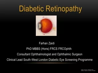
Diabetic Retinopathy • A condition that develops as a result of high blood sugar levels in both Type 1 and Type 2 diabetes. The blood vessels in the retina become weak, causing a varied degree of impact depending on the location and extent of the damage. May also result in cataracts (clouding of the lens) and glaucoma (increased pressure in the eye).
Diabetic Retinopathy VISUAL SYSTEMS IMPACTED • Blood vessels in the retina become damaged, potentially preventing the retina from properly changing light into nerve signals for the brain. • The vitreous (or middle of the eye) can be filled with scar tissue, blood and other fluid, causing pressure within the eye.
Diabetic Retinopathy EFFECTS ON VISUAL SYSTEM • Retinal detachment can occur, which would cause loss of vision or blurred vision. • Glaucoma (or increased pressure in the eye) can lead to blindness if not treated. • Cataracts (or clouding of the lens) can develop from the excess scar tissue or fluid in the eye, causing blurred vision.
Diabetic Retinopathy TREATMENTS • Treatments depend on the type, severity and response to previous treatments. • The nonproliferative (or non-progressive) type may only need to be monitored, while maintaining good blood pressure control. • The proliferative (or progressive) type requires surgical treatment.
Diabetic Retinopathy SURGICAL TREATMENTS • Focal laser treatment can stop or slow blood and fluid from leaking in the eye. During the procedure, abnormal blood vessels are treated with laser burns. • Scatter laser treatment can reduce the size of abnormal blood vessels. During the procedure, areas of the retina are treated with scattered laser burns. • Vitrectomy is a procedure that removes blood from the vitreous (or middle of the eye). During the procedure, the eye is given a small cut to remove scar tissue and blood.
Diabetic Retinopathy TESTING • Visual acuity testing – measuring the ability to focus • Ophthalmoscopy and slit lamp exam – detects changes in the retina and other structures • Gonioscopy – detects if the area where the fluid drains out of your eye is open or closed • Tonometry – measures the pressure of the eye which helps detect glaucoma
Diabetic Retinopathy MEDICATIONS • There are no approved medications that have been proven to prevent or even stop the progression of diabetic retinopathy. • Some medicines (such as insulin) have been determined to assist in preventing or delaying complications from diabetes.
Diabetic Retinopathy EXPERIMETAL MEDICATIONS • Ely Lilly & Co is testing a protein-based beta inhibitor (known as Kinase C-Beta) that is showing promise for preventing the progression of diabetic retinopathy. • Genetech is testing drugs for macular degeneration (loss of center vision) that may have future benefits for diabetic retinopathy.
Diabetic Retinopathy ONSET and OUTCOME • The condition is associated directly with another disease. It comes as a complication of both type 1 and type 2 diabetes. • The condition is generally progressive, but can be treated surgically to prevent or at least reduce the progression of the condition.
Diabetic Retinopathy PREVALENCE • 4.3% of people of all ages • 8.2% of people 40 years and older
Diabetic Retinopathy FUNCTIONAL IMPLICATIONS • Blurred vision or gradual vision loss can occur as a standard symptom of the condition. • Floaters (or tiny particles drifting across the eye) can increase in number and severity. • Shadows or missing areas of vision result from the presence of foreign material in the eye. • Difficulty seeing at night or in darkness can result when the retina fails to process light correctly.
Diabetic Retinopathy CARE • The main risk factors are poor nutrition and blood sugar maintenance • The most important care for Diabetic Retinopathy is having eye exams every 3 to 6 months in order to monitor for Glaucoma and Cataracts
Diabetic Retinopathy NATIONAL RESOURCE American Diabetes Association 1701 North Beauregard Street Alexandria, VA 22311-1717 1-800-342-2383 (National Headquarters) E-mail: AskADA@diabetes.org www.diabetes.org http://www.diabetes.org/
Diabetic Retinopathy STATE RESOURCE Texas Diabetes Council - Central Campus 1100 West 49th Street Austin, TX 78756 (512) 458-7111 http://www.dshs.state.tx.us/diabetes/
Diabetic Retinopathy AREA RESOURCE American Federation for the Blind - Texas 11030 Ables Lane Dallas, TX 75229-4524 (214) 352-7222 http://www.afb.org/
Diabetic Retinopathy LOCAL RESOURCE Ophthalmology Associates 1201 Summit Avenue Fort Worth, Texas, 76102 Tel: 817-332-2020 Email: info@fortworth2020.com http://www.fortworth2020.com/diabetic_retinopathy.aspx
Diabetic Retinopathy REFERENCES • http://bascompalmer.org/site/disease/disease_diabetic.asp • http://diabetes.webmd.com/tc/diabetic-retinopathy-medications • http://www.mayoclinic.org/retinal-diseases/diabetic-retinopathy.html • http://www.aoa.org/diabetic-retinopathy.xml • http://www.ncbi.nlm.nih.gov/pubmedhealth/PMH0002192/
Diabetic RetinopathyCase Study • On November 11, 2001, a 54 year old man had his annual diabetic visual evaluation. He had no complaints of his vision. He had stated that his blood sugar normally reads around 155mg/dl and on occasion he would get a reading of 220mg/dl. It was two years prior that he had his last eye exam. Records from this particular visit noted that the man had a diagnosis of mild non-proliferative diabetic retinopathy without clinically significant macular edema. All of this man’s ocular history was great except it was noted that he had hard exudates in the posterior pole, but away from foveal tissue. • His medical history included renal insufficiency, depression, hyperlipidemia, hypertension, cellulitis of the leg and type II diabetes with renal and ophthalmic manifestations. Medications include insulin injections b.i.d., dressings to treat cellulitis of the leg, Zestril (Lisin-opril, AstraZeneca) and Zocor (Simvastatin, Merck).
Diabetic RetinopathyCase Study • Entering his examination, his acuities were 20/40-2 OU and was best corrected at 20/25 OU. His tonometry showed that he had intraocular pressure of 12mm Hg OD and 15mm Hg OS. His results found no rubeosis, mild cataract development OU, and exudates and hemorrhages within 500 microns of the fovea OD with retinal thickening. Microaneurysms and hemorrhages were present in all four quadrants. This visit noted a diagnosis of mild/moderate non-proliferative diabetic retinopathy (NPDR) and clinically significant macular edema, also known as CSME (retinal swelling and cysts formation in the macular area). • A fluorescein study on March 4, 2002 showed scattered perfusion from microaneurysms O.S. greater than O.D. (see figure 2). The patient was treated with focal grid argon laser, with 27 burns O.D. and 57 O.S.
Diabetic RetinopathyCase Study • In September, 2002,six months later, the patient returned for a follow up visit and had entering acuities of 20/30 OU. The assessment included type II DM, moderate NPDR and minimal CSME O.U. The retinal practitioner believed that the problem, CSME OS was resolving so they rescheduled another follow up visit four months later. • Almost four months later, in January, 2003, the man returned for his visit and complained about blurry vision and reported that he had to stop driving. He also reported that his glucose levels were normal and measured 113mg that morning. The man said that after his last surgery, his vision was stable but could no longer find use in his glasses. Entering acuities were counting fingers at five feet OD and 20/60 OS. Diagnosis was altered to type II DM with severe NPDR and diffuse CSME OU. After further testing the same day, a marked increase in retinal edema was noted.
Diabetic RetinopathyCase Study • Five months later, in May, 2003, he returned for another follow up visit and received a laser treatment of 1400 burns OD and 1450 burns OS. • In July, 2003, his examination showed an entering acuity of bare light perception OU and proliferative changes and diagnosed type II DM with sever PDR OU were noted. • The practitioners questioned the dramatic change in this patient’s vision. This case illustrates why you must conduct an objective analysis of a patient's underlying medical condition. Upon careful review of this case, several important findings were noted. The most important was the patient’s glucose control over the past several years. The data on his glucose levels were collected and his Hemoglobin A1c counts. This analysis revealed the patient’s poor dietary compliance over several years.
Diabetic RetinopathyCase Study • A patient is considered a suspect for diabetes when fasting serum glucose levels reach between 100 and 140mg/dl. The diagnosis is likely when that number is over 140mg/dl. Further, the hemoglobin A1c count is also utilized. If elevated above 7.0, a positive diagnosis is likely. The patient had both of these extremely elevated over many years. When a practitioner takes this information into account, there becomes no point in ocular treatment option because the disease is already out of control. The patient needs to want to help themselves first.
Diabetic RetinopathyCase Study • This case study ends with suggestions to prevent and/or postpone vision loss. It also provides excellent visuals for a person to better understand the situation. Actual glucose/A1c levels should be available. Practitioner reports that patients can be easily misled by at-home readings which can often seem reasonable without considering the A1c levels from a lab. A1c counts are a better indication of a patient’s plasma glucose levels over the past three to four months. They provide a better indication of the patient’s compliance. Also, careful clinical examination is vital to monitor and treat ocular conditions. The practitioners say that patient education can never be overstated. When a patient realizes the severity of the situation and the implications that are held, it can help to motivate the patient to practice a higher form of control over the diabetes.
Diabetic Retinopathy REFERENCES • (Gibb & Olafsson) (Cassin, 2006, p. 87) (Levak, p. 132)Gibb, R., & Olafsson, H. (2006, September). Case study: Rapid progression of diabetic retinopathy. Retrieved from http://www.optometric.com/article.aspx?article=71734 • Cassin, B. (2006). Dictionary of eye terminology (p. 87). Gainesville, FL: Triad Publishing Company. • Levak, N. (n.d.). Low vision: A resource guide with adaptations for students with visual impairments (p. 132). Austin, TX: Texas School fo the Blind and Visually Impaired.