Download
1 / 18
350 likes | 1.32k Vues
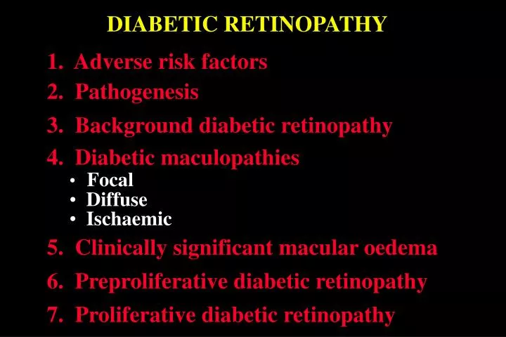
DIABETIC RETINOPATHY. 1. Adverse risk factors. 2. Pathogenesis. 3. Background diabetic retinopathy. 4. Diabetic maculopathies Focal Diffuse Ischaemic. 5. Clinically significant macular oedema. 6. Preproliferative diabetic retinopathy. 7. Proliferative diabetic retinopathy.

E N D
DIABETIC RETINOPATHY 1. Adverse risk factors 2. Pathogenesis 3. Background diabetic retinopathy • 4. Diabetic maculopathies • Focal • Diffuse • Ischaemic 5. Clinically significant macular oedema 6. Preproliferative diabetic retinopathy 7. Proliferative diabetic retinopathy
Adverse Risk Factors 1. Long duration of diabetes 2. Poor metabolic control 3. Pregnancy 4. Hypertension 5. Renal disease 6. Other • Obesity • Hyperlipidaemia • Smoking • Anaemia
Location of lesions in background diabetic retinopathy
Signs of background diabetic retinopathy Microaneurysmsusually temporal to fovea Intraretinal dot and blot haemorrhages Retinal oedemaseen as thickening on biomicroscopy Hard exudates frequently arranged in clumps or rings
Focal diabetic maculopathy • Focal leakage on FA • Circumscribed retinal thickening • Focal photocoagulation • Associated complete or incomplete • circinate hard exudates • Good prognosis
Diffuse diabetic maculopathy • Generalized leakage on FA • Diffuse retinal thickening • Grid photocoagulation • Frequent cystoid macular oedema • Guarded prognosis • Variable impairment of visual acuity
Ischaemic diabetic maculopathy • Macula appears relatively normal • Capillary non-perfusion on FA • Poor visual acuity • Treatment not appropriate
Clinically significant macular oedema Hard exudates within 500 m of centre of fovea with adjacent oedema which may be outside 500 m limit Retinal oedema within 500 m of centre of fovea Retinal oedema one disc area or larger any part of which is within one disc diameter (1500 m) of centre of fovea
Treatment of clinically significant macular oedema Grid treatment Focal treatment • For diffuse retinal thickening located more • than 500 mfrom centre of fovea and • 500 m from temporal margin of disc • For microaneurysms in centre of hard • exudate rings located 500-3000 m • from centre of fovea • Gentle whitening or darkening of • microaneurysm (100-200 m, 0.10 sec) • Gentle burns (100-200 m, 0.10 sec), • one burn width apart
Preproliferative diabetic retinopathy Signs • Dark blot haemorrhages • Intraretinal microvascular • abnormalities (IRMA) • Cotton-wool spots • Venous irregularities Treatment - not required but watch for proliferative disease
Proliferative diabetic retinopathy • Affects 5-10% of diabetics • IDD at increased risk (60% after 30 years) Neovascularization • Flat or elevated • Severity determined by comparing with area of disc Neovascularization of disc = NVD Neovascularization elsewhere = NVE
Indications for treatment of proliferative diabetic retinopathy NVE > 1/2 disc in area + haemorrhage NVD > 1/3 disc in area Less extensive NVD + haemorrhage
Laser panretinal photocoagulation • Area covered by complete PRP • Initial treatment is 2000-3000 burns • Spot size (200-500 m) depends • on contact lens magnification • Follow-up 4 to 8 weeks • Gentle intensity burn (0.10-0.05 sec)
Assessment after photocoagulation Poor involution Good involution • Persistent neovascularization • Regression of neovascularization • Haemorrhage • Residual ‘ghost’ vessels or • fibrous tissue • Re-treatment required • Disc pallor
Indications for vitreoretinal surgery Severe persistent vitreous haemorrhage Dense, persistent premacular haemorrhage Retinal detachment involving macula Progressive proliferation despite laser therapy