Download
1 / 40
440 likes | 717 Vues
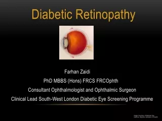
Diabetic Retinopathy. Steven Sanislo, M.D. Assistant Professor Stanford University Department of Ophthalmology. Diabetic Retinopathy. Diabetic retinopathy is a leading cause of new cases of blindness in people aged 20 to 74 years in the USA

E N D
Diabetic Retinopathy Steven Sanislo, M.D. Assistant Professor Stanford University Department of Ophthalmology
Diabetic Retinopathy • Diabetic retinopathy is a leading cause of new cases of blindness in people aged 20 to 74 years in the USA • Many of the complications of diabetic retinopathy can be prevented or delayed by blood glucose control and timely intervention.
Retinal Histology Sclera Choroid RPE Photoreceptor outer segments Photoreceptor inner segment Outer Plexiform layer Bipolar cells Inner plexiform layer Ganglion cells Nerve fiber layer
Retinal Diagnostic Tests • Fundus Photography • Fluorescein Angiography (FA) • Optical Coherence Tomography (OCT) • Ocular Ultrasonography • Electroretinography (ERG)
Pathogenesis of DR • Prolonged hyperglycemia is the major etiologic agent in all of the microvascular complications of diabetes, including diabetic retinopathy. • The cellular mechanisms through which hyperglycemia acts currently remain unclear
Pathogenesis of DR Mechanisms that have been proposed are: • 1. hyperglycemia may alter the expression of one or more genes, leading to increased (or decreased) amounts of certain gene products that can alter cellular functions. • 2. Glycosylated proteins can undergo a series of reactions, leading to considerable alteration of proteins. • 3. Chronic hyperglycemia may produce oxidative stress in cells, leading to the formation of an excess of "toxic end products of oxidation" including peroxides, superoxides, nitric oxide, and oxygen free radicals.
VEGF and DR • Vascular Endothelial Growth Factor • Promotes vascular growth and permeability • Elevated levels of circulating VEGF in conditions with retinal ischemia
Anatomic Changes Microanerysms • Damage to endothelial cells leads to dilated capillaries and venules • These altered vessels allow serum and blood to leak into the retina
Mechanisms of Vision Loss • Retinal ischemia • Macular edema • Vitreous hemorrhage • Epiretinal membrane formation • Retinal detachment • Neovascular glaucoma
Prevention • Prospective controlled interventional studies have shown that strict control of blood glucose and blood pressure significantly reduces and delays the onset and severity of diabetic retinopathy.
Screening Type 1 diabetics: First screen 5 years after onset, then annually. Type 2 diabetics: First screen upon diagnosis and then annually.
Treatment • NPDR without macular edema - • Observe • Macular edema - • 1. Focal/Grid laser photocoagulation • 2. Vitrectomy with membrane peeling • 3. Intraocular Steroid* • 4. Intraocular VEGF inhibitor* * Off-label use, contraversial
DME laser treatment * * * * * * * * * * * *
Treatment • Vitreous Hemorrhage - • 1. Pan-retinal photocoagulation • 2. Vitrectomy with laser photocoagulation • 3. Intraocular VEGF inhibitor* • Traction Retinal Detachment - • 1. Observation if not involving the macula • 2. Vitrectomy with membrane dissection * Off-label use, contraversial