Download

1 / 41
410 likes | 434 Vues
Chapter 3 DIAGNOSTIC RADIOLOGY Image quality Dr M A Oghabian Medical Physics Group, Tehran University of Medical Sciences www.oghabian.net. IAEA Post Graduate Educational Course Radiation Protection and Safe Use of Radiation Sources. Aim:

E N D
Chapter 3DIAGNOSTIC RADIOLOGYImage qualityDr M A OghabianMedical Physics Group, Tehran University of Medical Scienceswww.oghabian.net IAEA Post Graduate Educational Course Radiation Protection and Safe Use of Radiation Sources
Aim: To become familiar with the factors that determine the image clarity and the way the image quality can be improved IAEA Post Graduate Educational Course Radiation Protection and Safe Use of Radiation Sources
Contents • Image contrast • Blur or lack of sharpness • Distortion and Artifacts • Image noise Add module code number and lesson title
خصوصيات تصوير در راديولژي 1- دانسيته تصوير غيرخطي است. اما اين يك عدم مزيت نيست. • 2- تصویرداراي زمينه (Background) تشكيل شده از هر دو دانسيته و noise مي باشد. • -3داراي عدم وضوح(Unsharpness) بدليل focal spot size و Screen مي باشد. • -4داراي تغييرات فوتوني و نوري عمدتاً Random بنام noise مي باشد. Add module code number and lesson title
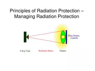
Factors affecting image quality Blurring or Unsharpness Contrast Image quality Distortion & artifact Noise Add module code number and lesson title
Image contrast • Difference in optical density between adjacent points on an image represents contrast • Contrast depends on: Density (air, bone) Thickness (micro calcifications) Atomic number kVp Film, intensifying screen, contrast agents Add module code number and lesson title
Receptor contrast • The film as receptor has a major role to play in altering the image contrast • There are high contrast and high sensitivity films • The characteristic curve of the film describes the intrinsic properties of the receptor • (base + fog, sensitivity, mean gradient, maximum optical density) • N.B.: Film processing strongly has a pronounced effect on fog and contrast Add module code number and lesson title
Video monitor • The video monitor is commonly used in fluoroscopy and digital imaging • The display on the monitor adds flexibility in the choice of image contrast • The dynamic range of the monitor is limited (limitation in displaying wide range of exposures) • Increased flexibility in displaying image contrast is achieved by adjustment of the window level or gray levels of a digital image Add module code number and lesson title
Blur or lack of sharpness • The boundaries of an organ or lesion may be very sharp but the image shows a lack of sharpness • Different factors may be responsible for such a degree of “fuzziness” or blurring • The radiologist viewing the image might express an opinion that the image lacks “detail” Add module code number and lesson title
Factors affecting image sharpness Subject Unsharpness Geometric Unsharpness Image Unsharpness Motion Unsharpness Receptor Unsharpness Add module code number and lesson title
Geometric blur • If the focal spot is effectively small, the blur is minimized because of minimal geometric unsharpness • As the focal spot increases, the blur in the image increases Large focal spot Small focal spot
Geometric Unsharpness • -مـحـوي مـربوط به اندازه نقطه كانوني با كاهش فـاصـله شئ تـا فـيـلـم يـا افـزايـش فـاصـلـة مـنـبـع تـشــعـشـع تــا شئ، كــاهــش مــي يــابـد. Add module code number and lesson title
Lack of sharpness in the subject • Not all structures in the body have well-defined boundaries (superimposition essentially present in most situations) • The organs do not have square or rectangular boundaries • The amount of required details in the object, is an essential requirement of any imaging system • The absence of sharpness, in the subject/object is reflected in the image
ِAbsorption unsharpness Add module code number and lesson title
Lack of sharpness due to motion (1) • Common and understandable blur in medical imaging • Patient movement : • uncooperative child • organ contraction or relaxation • heart beating, breathing etc. • Voluntary motion can be controlled by keeping examination time short and asking the patient to remain still during the examination
Lack of sharpness due to motion (2) • Shorter exposure times are achieved by the use of fast intensifying screens • N.B.: Faster screens result in loss of details (receptor sharpness) • Further, the use of shorter exposure time has to be compensated with increased mA to achieve a good image • This often implies use of large focal spot (geometric sharpness)
Movement Unsharpness • -مـحـوي مــربـوط بــه حـركــت بـيـمـار بــا كــاهــش زمــان تــصـويـربـرداري و يـا استـفـاده از آشـكـارسازهـاي سـريــع مـثل Cine- Fluorography مــي تـوانـد مـحــدود شـود • -از مـحـوي حـركـتـي جهت مـحـوكـردن بـخشهـايي از عـمـق بـافـت در تـومـوگــرافــي اسـتــفــاده مــيشـود Add module code number and lesson title
Lack of receptor sharpness • The intensifying screen in radiography has a crystal size which is larger than that of the emulsion on the film • An image obtained without the screen will be sharper than that obtained with the screen, but will require much more dose • The thickness of the screen further results in degradation of sharpness
Total Unsharpness 2/1 (Ua2 + Us2 + Um2 + Ug2 ) =Ut • در تــصــويـــربــرداري از اجســام بـــزرگ فـــقــط لبة ( edge) شئ تحت تأثير مــحــوي قـــابـــل تـشـخيـص نمی شود. • در تـــصـــويـــربـرداري از اجسـام كــوچـك (مــثـل مـــيـكـروكـلسيـفـيـكـيـشن و عروق كوچك - سنگ كوچك)،توزيع نور حاصل از محوي باعث از بين رفتن خصوصيات شكل شئ مي شود وً شدت و كنتراست تصوير نيزكاهش مي يابد. Add module code number and lesson title
Distortion and artifacts • Unequal magnification of various anatomical structures • Inability to give an accurate impression of the real size, shape and relative positions • Grid artifact (grid visualized on the film) • Light spot simulating micro-calcifications (dust on the screen) • Bad film screen contact, bad patient positioning
Magnification • از آنجائـيكه تـابش واگرا (Divergent) است،بـزرگـنمائـي تـصوير نـسبـت بـه شيئ ايـجـادمـي شـود. • جهت كاهش اثر بزرگنمائـي، فاصله شيء تا فيلم را كم كرده و لذا در مواردي مثل عكسبرداري از سينه، فاصله تيوب تا فيلم زياد مي شود. • همچنين طريقه قرارگرفتن بيمار (بطورAP يا PA) • بزرگنمائـي در بعضي مواقع مثل ماكروراديوگرافيتعمدا ايجاد مي شود. Add module code number and lesson title
Distortion • تغيير شكل ( Distortion) اجزا ناشي از زاويه متفاوت تابش و بـزرگــنمائـي حاصل مي شود. • جهت كاهش اثر تغيير شكل، فاصله شيء تا فيلم را كم كرده و بافت مورد تصويربرداري تا حد ممكن در مركز ميدان قرار داده مي شود. Add module code number and lesson title
Image noise • Information that is not useful is noise • The snowing in a TV image, the speckles in an ultrasound image are examples of noise • Noise interferes with visualization of image features useful for diagnosis
Main Sources of Noise Radiation noise (eg; heel effect) (1 2 - Random or Stochastic noise: • جذب Random فوتونهاي xدر صفحات تشديد كننده (Quantum noise). Photon Statistical Noise by low photon flux) (e.g; Screen or Coarse Grain Noise) جذب Random فوتونهاي نوريتوسط ذرات نقره در امولسيون فيلم Receptor or Fine Grain Noise Add module code number and lesson title
Main Sources of Noise • 2- Structured or nonstochastic: • - اختلاف در ضخامت مواد لايه تشديد كننده Screen باعث دانسيته غيريكنواخت فيلم میشود • Receptor noise, Non-uniformed film, screen defects,… • 4- Electronic noise : • زماني كه تقويت شديد سيگنال انجام مي شود باعث تقويت Random noise هم مي شود • 5- Random neural Process : • در چشم در زمان ديدن تصوير همچنين ايجاد نويز مي شود. • 6) Scattered Radiation in Both Subject and Imaging Receptors Add module code number and lesson title
Photon Statistical Noise (Quantum Mottle): • x-ray photons have a statistical character in nature • (the counts per unit time follows Gaussian distribution): • 1- One x-ray exposure is not identical to a second x-ray exposure • 2- The number of photons incident on different areas of the patient is not identical for a single exposure. • 3- Flow of x-ray energy is non-uniform • 4- There is a random variation in the number of photons striking each small area of the image receptor (Screen).
3. The Gaussian or Normal Distribution The Gaussian distribution is a probability distribution of a continuous random variable. If independent trails are conducted with an outcome having an arithmetic mean m and a standard deviation s, then
s=0.2 s=0.5 FWHM
Gaussian Distribution as a limiting case of the Poisson Distribution As n becomes large (eg; x-ray photon production) Poisson distribution becomes a Gaussian distribution. Since s 2 = m, This distribution has the properties of a Gaussian distribution: 68.3% of the events fall in the range of 95.5% 99.7%
Density Measurement Noise=s Noise and Detective Quantum Efficiency Noise is defined as the uncertainty in a signal due to random fluctuations in that signal. s 2s DD SNR = DD/ s Add module code number and lesson title
s s Noise is more prominent for low subject-contrast object
Signal-to -noise Ratio: Add module code number and lesson title
Signal to Noise Ratio Standard Deviation of a poisson distribution is: Where N represents the average number of photons striking a unit area. Most areas (68%) will then be struck by a number of photons in the range N- s to N+s. SNR ratio is the real measure of noise level. Quantum Noise Add module code number and lesson title
Noise Power Spectrum of film-screen system Add module code number and lesson title
Add module code number and lesson title 40 ناحيه A در اثر تغييرات ساختاري (structure)در سيستم تصويربرداري(مثل ضخامت screen و غير يكنواختي صفحه تشديد كننده يا غير يكنواختي تشعشع مي باشد. ناحيه B جايي كه نويز تقريباً ثابت است white noise ناميده مي شود و بدليل تغييرات اتفاقي مربوط به x-ray quantum يا نويز مدار الكتروني صورت مي پذيرد ناحيه C كه كاهش شديد نويز را نشان مي دهد بدليل خصوصيات transfer function است كه فركانس هاي در اين حد را با gain پايينتري انتقال ميدهد. ناحيه D(white noise), است مربوط به ذرات فركانس بالاي فيلم و اسكرين (grains) مي باشد.
Where to Get More Information • Hendee WR, Riternour ER, eds. Medical Imaging physics, 3rd ed. St. Louis: Mosby Year Book, 1992 • Sprawls Perry Jr. Ed. Pysical principles of medical imaging. Maddison: Medical Physics Publishing, 1993 • Moores BM, Wall BF, Eriskat H and Schibilla H, eds. Optimization of image quality and patient exposures in diagnostic radiology. London: British Institute of Radiology 1989. Add module code number and lesson title