Download
1 / 16
170 likes | 408 Vues
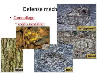
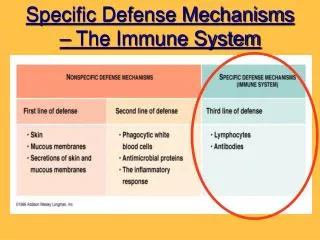
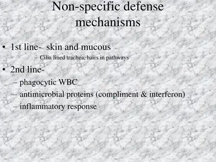
Non-specific defense mechanisms. 1st line- skin and mucous Cilia lined trachea, hairs in pathways 2nd line- phagocytic WBC antimicrobial proteins (compliment & interferon) inflammatory response. Phagocytic WBC. Neutrophils (60-70% of all WBC)

E N D
Non-specific defense mechanisms • 1st line- skin and mucous • Cilia lined trachea, hairs in pathways • 2nd line- • phagocytic WBC • antimicrobial proteins (compliment & interferon) • inflammatory response
Phagocytic WBC • Neutrophils (60-70% of all WBC) • attracted by chemical signals from damaged cells and enter tissues • Monocytes (5% of all WBC) • develop into macrophages which use psudopodia to capture invading bacteria • Eosinophils (1.5% of all WBC) • used to attack bigger invaders “worms” • natural killer cells • attack virus-infected cells to prevent spreading
Antimicrobial proteins • Compliment – various proteins in the plasma that work with antibodies, phagocytes, and on their own non-specifically to enhance immune response. • interferon-proteins secreted by virus-infected cells. Inhibits virus reproduction in neighboring cells. Can be mass produced now and may be used to treat cancer patients.
Inflammatory response • 1. Damaged tissues release chemical signals such as histamine (contained in basophils WBC and mast cells of connective tissue) and prostaglandins to increase blood flow. • 2. Prostaglandins induce vasodilation and increased permeability to clotting factors. • 3. Chemokines release chemicals that mediate the arrival of phagocytic cells to the area • Phagocytes consume debris and pathogens forming pus • Sometimes allergies cause massive release of histamine to “safe” invaders, so antihistamines block this response
Specific defense (3rd line) • Four major features • Specificity (recognize particular antigens) • diversity (responds to millions of different invaders) • self/nonself recognition- • memory - acquired immunity so the second time body is infected the response will be quick enough to avoid serious infection. This is the basis for vaccination.
Cell surface markers • Blood cells A, B, and Rh Factor proteins • Major histocompatibility complex (MHC) are glycoproteins marking cell as self • MHC class I are on all nucleated cells • MHC class II are only on specific immune cells • These allow cytotoxic T-cells (MHC I) and helper T-cells (MHC II with antigen fragments attached) to bond to cells • Huge amount of variety, so each individual is unique in their MHC proteins.
Humoral immunity • Results in production of antibodies • Free antigens activate B- cells • B- cells make the antibodies and then develop into Plasma cells and memory B-cells for next time • Plasma cells secrete antibodies • antibodies attach to antigens making them easy “prey” for phagocytes and complement
Formation of lymphocytes • Lymphocytes are WBC formed in the bone marrow. (B and T cells) • B- cells fully develop in the bone marrow before being released • T - cells then travel to the the thymus for further development before leaving. • In the thymus they pick up recognition of MHC complex as self.
Cell-mediated immune response • Antigens displayed by MHC class I glycoproteins in infected cells activate Cytotoxic T cells • Cytotoxic T cell give rise to memory T cells and Active cytotoxic T cells • Activated cytotoxic T cells attack cells by binding to and lysing them
2nd exposure • 2nd time the antigen stimulates Memory B cells and memory T cells to activate both humoral and cell-mediated responses. • 2nd defense (about 3 days) where as 1st response is usually 7-10 days. • Supressor T cells are thought to help turn off the immune response when antigens are gone.
Antibodies • A class of proteins referred to as the immunoglobulins (Ig) with 4 polypeptides (2 heavy chains and 2 light chains) held together by disulfide bridges to give them their quaternary structure • Most of the antibody structure is identical for all antibodies with the “tips” variable that bind to the epitope (exposed) region on the antigen surface. • Five types of immunoglobulins are divided by their constant regions IgM, IgG, IgA, IgD, and IgE.
Immunoglobulin classes • IgM: large molecule that initiates response by agglutinating (clumping up) the antigens • IgG: Most plentiful, triggers complement proteins • IgA: Prevents attachment of antigens to epithelial linings. Plentiful in mucus. • IgD: found on B-cells and probably initiate the development of B-cells into plasma cells • IgE: small number, trigger the histamine release of basophils and mast cells via receptor binding.
Helper T-cells • Helper T cells are stimulated by interleukin 1 of macrophages after engulfing antigens and presenting them. • Helper T cells in turn stimulate the B cells of the humoral defense and the Cytotoxic T cells of the Cell mediated defense by releasing interleukin 2.