Download
1 / 69
1.04k likes | 2.45k Vues
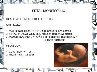
Fetal Heart Rate Monitoring. Paul G. Tomich , M.D. Department of Obstetrics and Gynecology University of Nebraska College of Medicine . Learning Objectives. Evolution Examples Descriptions Reassuring patterns Concerning patterns Definitions of Category I, II, and III tracings

E N D
Fetal Heart Rate Monitoring Paul G. Tomich, M.D. Department of Obstetrics and Gynecology University of Nebraska College of Medicine
Learning Objectives • Evolution • Examples • Descriptions • Reassuring patterns • Concerning patterns • Definitions of Category I, II, and III tracings • Discuss action needed • Non-stress Test (NST) • Biophysical Profile (BPP)
“Evolution” of FHR Monitoring • Monitoring fetus in labor • FHR patterns • Good outcomes • Poor outcomes • Contraction Stress Test (CST) • Non Stress Test (NST) • Biophysical profile (BPP) • Categorization of FHR Tracing into Category I, II, and III
Categorization of FHR Tracings • Recommendation of three-tiered system • April 2008 • More standardized interpretation • Concept: Interpretation of a FHR monitor strip is a dynamic process, with determination of whether a particular strip is reassuring and what action plans should be taken… and then to evaluate at a later time
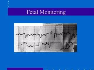
Ways to Monitor Uterine contractions Fetal heart rate (FHR)
Ways to Monitor Uterine contractions Fetal heart rate (FHR)
Features to Describe • Fetal heart rate (FHR) • Top line on monitor strip • Uterine contractions • Bottom line on monitor strip
Features to Describe Baseline Variability Accelerations Decelerations Trends over time Interpret into 1 of 3 categories
Baseline • Mean fetal heart rate • Rounded to increments of 5 • During a 10 minute period • Excluding accelerations and decelerations • Normal baseline • 100-160 BPM
Baseline • Bradycardia <100 BPM • Tachycardia >160 BPM • Indeterminate • less than 2 minutes of baseline is present
Fetal Tachycardia • Normal variant • prematurity • Intra-amniotic infection • Fetal anemia • Fetal cardiac arrhythmia (SVT) • Fetal hypoxia
Features to Describe Baseline Variability Accelerations Decelerations Trends over time Interpret into 1 of 3 categories
Variability • Fluctuations in FHR • Over 10 minutes • Descriptors are: • Absent: undetectable amplitude range • Minimal: undetectable up to 5 BPM • Moderate: amplitude range 6 to 25 BPM • Marked: amplitude range greater than 25 BPM
Features to Describe Baseline Variability Accelerations Decelerations Trends over time Interpret into 1 of 3 categories
Accelerations • Abrupt increase in FHR • At least 15 BPM above baseline • Duration • Must last 15 seconds to 2 minutes • Prolonged accelerations • Last 2 minutes to 10 minutes • Baseline change • Acceleration lasting 10 mins or longer
>15 beats above baseline 15 seconds to 2 minutes in length
Features to Describe Baseline Variability Accelerations Decelerations Trends over time Interpret into 1 of 3 categories
Decelerations • Decrease in baseline • 3 Types • Early • Variable • Late
Deceleration Decrease in FHR
Early Deceleration • Symmetrical to contraction • Mirror image of contraction • Gradual decrease in FHR • 30 secs or more from onset to nadir
EARLY DECELERATION • Gradual FHR decrease • Onset to nadir 30 seconds or more • Nadir of deceleration occurs with peak of contraction • Mirror contraction
Late Decelerations • Deceleration is delayed in timing • Occurs after the contraction • A gradual FHR decrease • Onset to nadir > 30 second
Variable Decelerations • Abrupt decrease in fetal heart rate • Onset to nadir less than 30 seconds • Decrease in FHR • 15 BPM or more • Lasting 15 seconds to 2 mins
VariableDeclerations • Pathophysiology • umbilical cord compression
Decelerations • Prolonged deceleration • Decrease of 15 BPM • Lasts 2-10 minutes • Baseline change • Deceleration lasting at least 10 mins • Description • Intermittent • Less than 50% of contractions in 20 minutes • Recurrent • More than 50 % of contractions in 20 minutes
Sinusoidal Pattern Smooth sin-wave pattern Cycle frequency 3-5 mins Persists for 20 minutes or longer
Uterine Contractions • Number of contractions in 10 minutes • averaged over thirty minutes • Document • Frequency • Intensity • Duration • Relaxation • time between contractions
Tachysystole • >5 contractions in 10 mins • Averaged over 30 mins
Categorization of FHR Patterns An evaluation of the fetus at a particular point in time Categories I, II, and III