Download
1 / 25
320 likes | 342 Vues
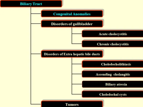
SOL Liver. Common liver lesions. Approach to liver SOL. History Clinical examination Lab – Hemogram, LFT, Albumin,INR Serology – ALA , Hydatid Tumor markers – AFP,CEA, CA 19-9. Presentation. Asymptomatic Nodule on screening in cirrhotic patients Presenting with pain and fever

E N D
Approach to liver SOL • History • Clinical examination • Lab – Hemogram, LFT, Albumin,INR • Serology – ALA , Hydatid • Tumor markers – AFP,CEA, CA 19-9
Presentation • Asymptomatic • Nodule on screening in cirrhotic patients • Presenting with pain and fever • SOL in a known patient of extrahepatic malignancy
Algorithm for Solitary SOL of Liver on USG Nagral S , Clinics in GI Surgery
Algorithm for Solid lesions on USG Nagral S , Clinics in GI Surgery
Algorithm for Multiple Liver SOLs on USG Nagral S , Clinics in GI Surgery
Beyond USG – CT • Symptomatic lesions – no response to treatment • Alterations in LFT • Underlying liver disease
When MRI ----- HCC vs regenerating nodule vs dysplastic nodule atypical lesions
Role of Biopsy Usually not required Diagnostic uncertainty
HISTORY • 54 yrs gentleman, no comorbidities • Frequency, urgency, in • complete evacuation • No other GI symptoms • USG abdomen – hypo echoic liver lesions • Lab - normal
Colonoscopy • Ulcero proliferative lesion in sigmoid colon • No synchronous lesions/polyps • Biopsy - adenocarcinoma
Chemotherapy • FOLFOX + bevaxizumab – 7 cycles • FOLFOX – 5 cycles
Surgery • Sigmoid colectomy + left lateral segmentectomy + metastectomy (seg 4b, seg 5, seg 7)