Download

1 / 12
120 likes | 186 Vues
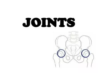
Joints. Chapter 8. Classifying Joints. Functions Flexibility for movement Hold bones together Structural Classification (binding tissue and cavity) Fibrous Cartilaginous Synovial Functional Classification (amount of movement) Synarthroses – immovable; axial skeleton

E N D
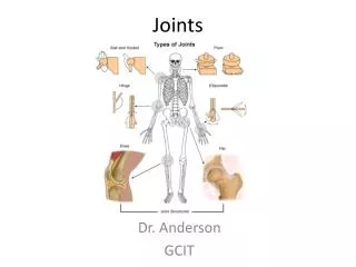
Joints Chapter 8
Classifying Joints • Functions • Flexibility for movement • Hold bones together • Structural Classification (binding tissue and cavity) • Fibrous • Cartilaginous • Synovial • Functional Classification (amount of movement) • Synarthroses – immovable; axial skeleton • Amphiarthroses - slightly movable; axial skeleton • Diathroses - freely moveable; limbs
Fibrous Joints • Sutures • Only b/w skull bones • Bind, but allow growth • Complete as an adult (synostoses) • Syndemoses • Longer than sutures • Length determines mov’t* • Interosseous membrane and tibia-fibula distal ends • Gomphoses • Tooth in alveolar socket • Periodontal ligament
Cartilaginous Joints • Synchondroses • Temporary, become synostoses • Epiphyseal plates and coastal cartilage • Symphyses • Cartilage fused to fibrocartilage pad/plate • Strength with flexibility • Intervetevbral discs and pubic symphysis
Synovial Joints • Articular cartilage • Joint (synovial) cavity • Articular capsule • Fibrous capsule (ext-) DICT • Synovial membrane (int-) LCT • Synovial fluid • Viscous, but thins with mov’t • Reinforcing ligaments • Double jointed = looser/stretchierligmanents and capsule • Rich blood and nerve innervation • Fatty pads for cushioning (hip and knee) • Menisci divide cavity and improve fit (knee and jaw)
Synovial Joints • Preventing friction • Bursae are flattend fibrous synovial sacs • Tendon sheaths are elongated bursa wrapped around a tendon • Stability • Articular surface shape • Determine mov’t & some stability • Ligaments • Prevent excessive/undesirable mov’t • Inadequate than stay stretched (taffy) = snapping • Muscle tone • Tendons stay taut so reactive
Joint Articular Shapes • Plane - articular surface is flat, nonaxial • Intercarpal and –tarsals; slip 1 or 2 ways • Hinge – cylindrical projection to a trough • Elbow and interphalengeal; 1 plane of mov’t • Pivot – rounded end into ring or “sleeve” • C1 & C2 or radius & ulna; 1 plane of mov’t • Condyloid - oval surface into a depression • Metacarpophalangeal (knucles); 2 planes of mov’t • Saddle – concave and convex surface • Carpometacarpal thumb joint; 2 planes of mov’t • Ball and socket – spherical end with a cup-like socket • Shoulder or hip joint; 3 planes mov’t
Synovial Movements • Gliding • Slips surfaces across one another • Flexion/extension • Reduces angle of joint/ increases angle • Abduction/adduction • Away from center/ toward midline • Pronation/supination • Face or palm down/ face or palm up • Rotation/circumduction • Turning on an axis/ making small circles • Inversion/eversion • Turn sole medially/ turn sole laterally • Dorsiflexion/plantar flextion • Flex/ point • Protraction/retraction • Jaw out/jaw in • Elevation/depression • Lift superiorly/move inferiorly
Knee Joint • Single cavity w/ 3 joints • Capsule partially encloses • Strong vertical force, weak lateral • Patellar ligament (knee-jerk) • Prevent hyperextension • Fibular and tibial collateral ligaments • Prevent lateral and medial rotation w/ extension • Anterior and posterior cruciate ligaments (tibial attach) • Prevent forward sliding of tibia; back displacement of tibia or forward femur • Posteriorly, medially, up; anteriorly, laterally, up • Lateral and medial meniscus anterior cruciate ligament
Temporomandibular Joint (TMJ) • Mandibular condyle is egg-shaped • Temporal bone is knob forward, concave fossa • Side-to-side is lateral exclusion, unique to mammals • Shallow socket = easy dislocation • Reset: thumbs in molars, push inferior and posterior • Pain from muscles tension
Clinical Terms • Sprain: stretching/tearing of a ligament • Dislocation (luxation): bones forced out of position • Bursitis: inflammation on bursa; blow or friction • Arthritis: synovial membrane thickens, production decrease • Osteoarthritis – degenerative; tissue thickens & bone spurs formed • Rheumatoid arthritis - autoimmune • Gouty arthritis – uric acid accumulation in soft tissue joints • Synovitis: inflammation of synovial membrane • Tendinitis: inflammation of tendon sheaths, overuse;