Download
1 / 8
80 likes | 190 Vues
Cell Suicide in Health and Disease. Keith McInerney Melissa Snyder Bob Aquavia. Characteristics of Apoptosis. (cell suicide) through the activation of a genetic program usually affects individual cells and its tissue distribution is patchy and asynchronous

E N D
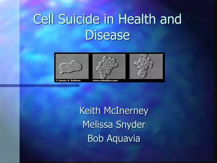
Cell Suicide in Health and Disease Keith McInerney Melissa Snyder Bob Aquavia
Characteristics of Apoptosis • (cell suicide) through the activation of a genetic program • usually affects individual cells and its tissue distribution is patchy and asynchronous • After only one day, they "commit suicide" and are replaced by younger cells. • cells break into smaller pieces called apoptotic bodies that other body cells recognize and eat.
BCL-2 • A proto-oncogene activated by chromosome translocation in B cell lymphomas • Encodes for a plasma membrane protein that inhibits apoptosis. • Appears localized or associated with intracellular membranes of mitochondria the ER, and nuclei • Regulation of the redox potential of the cell, • Suppresses cell death induced by oxidizing agents and appears to affect glutathione levels (Korsmeyer et al., 1993; Zhong et al., 1993).
p53 • protien that is mutated in more that 50% of tumors • normal form of p53 keeps the cell from entering cell division cycle • found to bring about apoptosis after DNA damage has occurred.
FAS ligand • Fas ligand expression by cancer cells induce apoptosis of activated T cells and contribute to immune tolerance. • never been explored in vivo in tumor cell models yielding either immune response or tolerance. • When it binds to Fas on the same cell or on another activated T cell at the site of infection, the cell is instructed to undergo apoptosis.
FAS • Resting T cells produce low levels of FAS which spans the cell membrane. • It projects into the extracellular apace at one end and into the cell’s interior at the other end. Here it can convey signals deeper into the cell. When T cells first encounter an antigen, they make extra, but non-functional FAS. Later, the FAS binds to the FAS ligand in order to facilitate apoptosis.
ICE-like Proteins • Called this because they structurally resemble interleukin-1 converting enzyme • protein-cleaving enzyme • when enzymes are activated, they chop various other proteins in ways that lead to destruction of the cell
THE END All of these proteins contribute to the regulation and outcome of apoptosis.