Download
1 / 29
290 likes | 873 Vues
Neurological Examination of Spinal Cord injury. Dr. Osama Neyaz Assistant Professor Department Of PMR. Anatomy of spine. 7 cervical vertebrae 12 thoracic vertebrae 5 lumbar vertebrae 5 fused sacral vertebrae 3-4 small bones comprising the coccyx

E N D
Neurological Examination of Spinal Cord injury Dr. Osama Neyaz Assistant Professor Department Of PMR
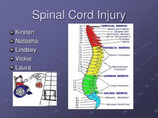
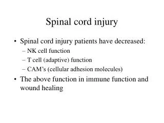
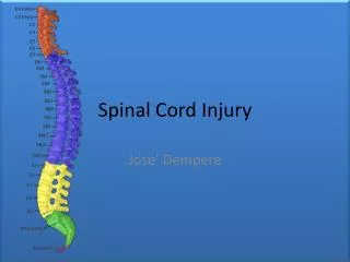
Anatomy of spine • 7 cervical vertebrae • 12 thoracic vertebrae • 5 lumbar vertebrae • 5 fused sacral vertebrae • 3-4 small bones comprising the coccyx • Spinal cord ends as conus medullaris at level of first lumbar vertebra lumbar and sacral nerve roots exit below this and form the cauda equina
Neuroanatomy • 1&2 Posterior Columns: convey Ipsilateral information about two Point discrimination, proprioception And vibratory sense • 5 Lateral Spinothalamic Tract: carries Pain and Temperature Information From contralateral extremity • 4 Lateral Corticospinal Tract: Carries Motor Information from Contralateral Brain to Ipsilateral Extremity
Mechanisms of Injury • Compression • Flexion Injury • Extension Injury • Rotation
Compression Injury • Vertebral body fracture • Disc herniation • Epidural hematoma • Displacement of posterior wall of the vertebral body
Jefferson Fracture • A comminuted fracture of the ring of C1. • Compression of base of skull against C1 • Results in cracking the ring of C1 • Best seen on open mouth x-ray
Atlantoaxial and Dens Fractures • The result of hyperflexion or hyperextension injuries 8% of Dens Fractures associated with C1 fractures C2 Fractures Dens Fracture : • HyperflexionInjury Hangman Fracture : • Hyperextension Injury • Traumatic spondylolisthesis of the axis • Bilateral fractures through the pars interarticularis of the axis
flexion teardrop fracture • Hyperflexion of the subaxial cervical spine • Retropulsion of the larger portion of a vertebral body into the spinal canal, detached from an anterior fragment (teardrop) • Often associated with an anterior cord syndrome.
clay-shoveler’sfracture • Avulsion fracture of the spinous process of C6, C7, or T1. • It is not typically associated with neurologic injury.
Thoracolumbar Trauma • Mechanism of injury • Compression • Distraction • Rotation
Chance Fracture • Failure of all three columns due to flexion-distraction • Falls from a height • Strikes part of the torso on an immovable object • Injury pattern most likely to cause SCI
The three-column concept of spinal anatomy • The anterior column: ALL + anterior portion of the vertebral body+ anterior portion of the disk. • The middle column: posterior portion of the vertebral body + the posterior portion of the disk + PLL • The posterior column: the pedicles facet joints + laminae + supraspinous ligament, interspinous ligament + facet joint capsule + ligamentumflavum.
Stable Vs UNSTABLE FRACTURE • When the integrity of the middle and either the anterior or the posterior column is affected, the spine is likely to be unstable. • The columns can be affected by: • Fracture • Ligamentous disruption • Gunshot wounds • Because of the nature of the injury, can affect more than one column and the spine can still remain stable. • SCI can occur without obvious radiographic findings.
Clinical Syndromes after Incomplete Spinal Cord Injury • Central Cord Syndrome • Brown-SequardSyndrome • Anterior Cord Syndrome • Conus Medullaris Syndrome • Cauda Equina Syndrome
Central Cord Syndrome • Motor>Sensory Loss • Upper>Lower Extremity Loss • Distal >Proximal Muscle Weakness • Classically occurs with hyperextension injuries of the cervical spine
Brown-Sequard Lesion • A burst fracture with posterior displacement of bone fragments compresses one side of the spinal cord. • Loss of Ipsilateral Proprioception, Light Touch and Motor Function • Loss of Contralateral Pain and Temperature Sensation • Due to hemisection of the cord due to penetrating injury • Incomplete lesions most common
Anterior Cord Syndrome • A large disk herniation compresses the anterior aspect of the spinal cord, leaving the dorsal columns intact. • Loss of Motor function, Pain and Temperature Sensation • Preservation of Light touch, Vibratory Sensation and Proprioception
Conus Medullaris Syndrome A burst fracture of with posterior displacement of bone fragments compresses the conus medullaris. Injury to sacral cord, lumbar nerve roots causing • Areflexicbladder • Loss of control of bowels • Knee jerk relexes preserved, ankle jerk absent • Signs similar to cauda equina syndrome except more likely to be bilateral
Cauda Equina Syndrome • A central disk herniation at L4-L5 level compresses the cauda equina. • Injury to nerve roots and not spinal cord itself • Muscle weakness and decreased sensation in affected dermatomes • Decreased bowel and bladder control
Classification of Spinal Cord Injury Patients are classified according to the ASIA Impairment Scale (AIS) • Combined efforts from • ASIA: American Spinal Injury Association • ISCOS: International Spinal Cord Society
Components of the Test Three Main Parts to the Exam: • Strength Testing • Light Touch Sensation • Pinprick Sensation Lowest Level of motor control: • Voluntary Anal Contraction • Lowest Level of Sensation: • Deep Anal Pressure
Neurologic Exam: Dermatomes • C5- Deltoid • C6 – Thumb • C7 – Middle Finger • C8 - Little Finger • T4 – Nipple • T8 – Xiphoid • T10 - Umbilicus • T12 – Symphysis Pubis • L4 – Medial aspect of leg • L5 - Space between first and second toes • S1 – Lateral border of the foot • S3 – Ischial Tuberosity • S4-5 – Perianal region
Myotomes • C5 – Deltoid • C6 – Wrist Extensors • C7 – Elbow Extensor • C8 – Finger flexors • T1 – Little finger abduction • L2 - Hip flexion • L3 - Knee Extension • L4 - Ankle dorsiflexion • L5 - Toe extension • S1 – Plantar flexion