Download
1 / 24
250 likes | 444 Vues
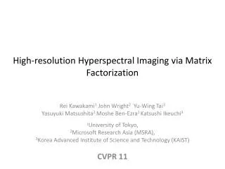
High Resolution imaging in Deep Tissue. Rainer Heintzmann , Institute for Photonic Technologies (IPHT), Friedrich Schiller University of Jena Randall Division, King‘s College London. Biophotonics, 2011. The Problem: 1. Absorbtion. blood. melanosom. Absorbtion Coefficient / cm -1.

E N D
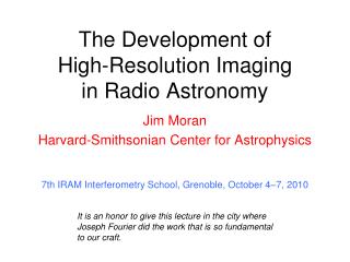
High Resolution imaging in Deep Tissue Rainer Heintzmann, Institute forPhotonic Technologies (IPHT), Friedrich Schiller University of Jena Randall Division, King‘s College London Biophotonics, 2011
The Problem: 1. Absorbtion blood melanosom Absorbtion Coefficient / cm-1 aorta water skin epidermis Wavelength / nm
User requirements • as „lifelike“ as possible: • the illumination light should not influence the behaviour • sample-mounting shound not disturbe • fluorphores (vFPs) should behave close towildtype • images have to be taken quickly„temporal sampling“to avoid artefacts, distortions and allow tracking
The Problem: 2. Scattering Rayleigh scattering: ~ l-4 Blue: Bad! Red / Infrared: OK!
focal distance plane of focus Physics of Light Light as a Wave Moving Phase Front
The Problem: 3. Aberrations Distorted Wavefront (Aberrated)
Solution 1: Two Photon Effect ps ps ps ps hn hn hn Singlet Singlet Ps1 Ps1 Ps0 Ps0 2 Photons absorbed 1 Photon absorbed Probability ~ Intensity2
Solution 1: Two Photon Effect Two Photon Excitation Zipfel, Williams, Webb, Nature Biotechnology21, 1369 - 1377 (2003)
Solution 1: Two Photon Effect DichromaticReflector Wide Area Detector at close destance emission photons will be multiply scattered Non descanned detection needed to maximize capture area
Solution 1: Two Photon Effect • Much less absorption • Much less scattering • Less aberrations • Less out-of-focus bleach • Fancy technique: Temporal focussing leads to ultrafast scans Scattering loss is compensated until Surface starts „burning“
Solution 1: Two Photon Effect Some techniques (Multifocus, Temporal focussing) require imaging of the emitted light Problem: haze from emission scattering remains Solution: 1. Use different technique (e.g. single beam) 2. Combination with modulated excitation e.g. Structured illumination or focal modulation removes haze, but subtraction noise remains 3. temporal gating (may reduce noise but also signal)
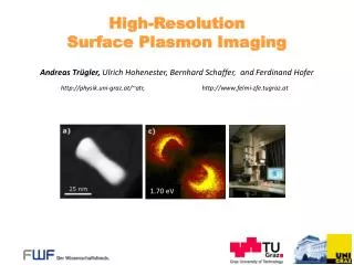
Solution 2: Ultramicroscopy Unnessesary Bleaching Selective Plane Illumination Microscopy Illumination Plane of focus
Solution 2: Ultramicroscopy Selective Plane Illumination Microscopy Light Sheet Illumination Cylinder Lens
Solution 2: Ultramicroscopy Selective Plane Illumination Microscopy Detection Illumination
Solution 2: Ultramicroscopy 3D reconstruction of large 3-dimensional microscopic systems • Material above or below the focal plane are not illuminated no out-of-focus blur & no out-of-focus bleaching • Simple collection optics images illuminated area onto a camera • Rotation different angles of view 3D tomographic image reconstruction
Solution 2: Ultramicroscopy 3D reconstruction Mouse embryo E12.5. (A) Surface (B) Stained nerve fibers (C) Surface of head(D) Sensory nerve fibers innervating the vibrissae. Because the mouse embryos are opaque, a special clearing technique is applied: Index matching:2 parts benzyl benzoate and one part benzyl alcohol).
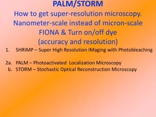
Overview: Direct Imaging • Stimulated Emission Depletion Microscopy • Pointillism, PALM and STORM • Selective Plane Illumination Microscopy • Structured Illumination • Circumventing the limit: Nonlinearity • Interferometric Resolution Enhancement
x,y a z NA = n sin a Problem: Limited Numerical Aperture Objective Lense Immersion Medium Cover Slip Immersion Medium Cell Slide
Aperture increaseby rotation of the specimen x,y z Solution: Axial Tomography Objective Lense Immersion Medium Cover Slip Immersion Medium Cell Glass Fiber Shaw et al., Cogswell et al.,Kawata et al., Heintzmann et al.
Reconstructed Estimate View 1 View 2 Apply Measured Convolution Register , , Back Convolution View 1 View 2 C NC Ei Simulated Mi Combined ML-Deconvolution Compare
Biological Specimen Moss Spore Polytrichum Commune Fully automatically registered R. Heintzmann and C. Cremer., J. Microsc., 206 (1),7-23, 2002
Biological Specimen Moss Spore Fully automatically registered Polytrichum Commune
Solution 2: Ultramicroscopy • Much lessout-of-focus bleach • complicated sample mount • Absorption/scatteringlosscanbe a problem(excitationandemisission) • combinations (2-photon excitation) withstructuredilluminationorsweptlineilluminationarepossible
Solution 3: Aberration Correction Pre-compensation element (SLM) • Quality improvement is often only moderate • Estimation of aberrations is difficult and time consuming(Selective Aperture, iterative procedure, ..) • spatially varying aberrations are very hard