Download
1 / 1
40 likes | 326 Vues
Photoactivation Localization Microscopy (PALM) in Biological Samples Chih -Jung Hsu, Janis Burkhardt and Tobias Baumgart , NSEC DMR08-32802.

E N D
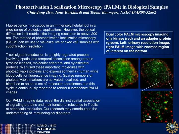
Photoactivation Localization Microscopy (PALM) in Biological Samples Chih-Jung Hsu, Janis Burkhardt and Tobias Baumgart, NSEC DMR08-32802 Fluorescence microscopy in an immensely helpful tool in a wide range of biological applications. However, the optical diffraction limit restricts the imaging resolution to above 200 nm. The method of photoactivation localization microscopy (PALM) can be use to visualize live or fixed cell samples with subdiffraction resolution. T-cell signal transduction is a highly regulated process involving spatial and temporal association among protein tyrosine kinases, molecular adaptors, and cytoskeletal proteins. We fused these important molecules with photoactivable proteins and expressed them in human white blood cells for fluorescence imaging. Sparse numbers of photoactivable markers are activated, localized, and bleached to obtain a set of molecular coordinates and this cycle is continuously repeated to render fluorescence PALM images. Our PALM imaging data reveal the distinct spatial association of signaling proteins and their functional relevance in T cells at nanoscale resolution. Our research may contribute to the understanding of immunological disorders. Dual color PALM microscopy imaging of a kinase (red) and an adapter protein (green). Left: orinary resolution image, right PALM image with zoomed region of interest on the bottom. 5μm 1μm