Download
1 / 59
600 likes | 738 Vues
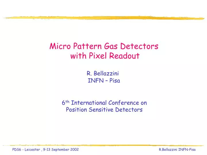
Micro Pattern Gas Detectors with Pixel Readout R. Bellazzini INFN – Pisa. 6 th International Conference on Position Sensitive Detectors. The MSGC. Cross-section of a MSGC. Simulated current signals. Electric field lines strongly concentrates on anode strips.

E N D
Micro Pattern Gas Detectors with Pixel Readout R. Bellazzini INFN – Pisa 6th International Conference on Position Sensitive Detectors
The MSGC Cross-section of a MSGC Simulated current signals Electric field lines strongly concentrates on anode strips Signals 10 times faster with respect to MWPC signals
Rate capability From MWPC To MSGC Rate capability increases 3 orders of magnitude Rate capability > 1 MHz/mm2 R. Bellazzini et al., Nucl. Instr. Meth.
The MicroGap Chamber (MGC) The space charge effect, in the MSGC, is strictly connected to the presence of an insulating substrate. When a significant fraction of ions of the avalanche hits this surface, it causes a charge-up which reduces locally the electric field. In the MGC the insulating surface exposed to the gas is strongly reduced. Scanning electron microscope picture R. Bellazzini et al., Nucl. Instr. Meth. A335 (1993) 69 Electric field lines close to the anodes are even more intense than in a MSGC resulting in a very fast signal development.
MWPC MSGC MGC Signal evolution from MWPC to MGC Signals Electric field
MGC performances Short term measurement of gain stability The MGC can withstand high rate of radiation (up to few MHz/mm2 ) without visible change in gain. No evidence of charging-up effect with time is observed
Linearity of GEM current at very high counting rates. Counting rate linear up to 2 MHz/pixel (limited by electronics dead-time) D. Pacella et al., Review of Scientific Instrument, Vol.72 No.2 (2001)
The Gas Electron Multiplier (GEM) A new class of position sensitive MicroPattern Gas Detectors, robust and cheap, has been developed using the advanced printed circuit (PCB ) technology: GEM, Micro Groove and Well Detectors The GEM is a thin polymer foil (Kapton), metal coated on both sides, chemically pierced by a high density of holes. On application of a voltage gradient, electrons released on the top side drift into the hole, multiply in avalanche and transfer to the other side. Proportional gains above 103 are obtained in most common gases. F. Sauli, Nucl. Instr. Meth. A386 (1997) 531
Cu-plated Kapton Copper etching Kapton etching Edge finish GEM manufacturing Typical geometry: 5 mm Cu on 50 mm Kapton 70 mm hole at 140 mm pitch
The GEM Charge amplification and read-out take place on separate electrodes. The read-out PC board can be structered in a multi-pixel pattern to get full 2D imaging capability. R.Bouclier et al., Nucl. Instr. Meth. A396 (1997) 50
Fast trigger Y-coordinate X-coordinate Two-dimensional Readout The electron charge is collected on strips or pads on the read-out board. A fast signal can be detected on the lower GEM electrode for triggering or energy discrimination. Cartesian Small angle Pads
Full width 20 ns (2 mm gap) The GEM signal The duration of the signal correspond to the drift time of the electrons in the transfer gap.
GEM + Pixel Read-Out: recent applications The complete separation between the amplification structure and the pick-up electrodes allows full flexibility on the choice of read-out pattern GEM+Micro Pixel electrode Recent applications: X-ray Polarimetry Time resolved plasma diagnostic
X-ray Astrophysical Polarimetry Polarization from celestial sources may derive from: • Emission processes themselves: cyclotron, synchrotron, non-thermal bremmstrahlung. • (Westfold, 1959; Gnedin & Sunyaev, 1974; Rees, 1975) • Scattering on aspherical accreting plasmas: disks, blobs, columns. • (Rees, 1975; Sunyaev & Titarchuk, 1985; Mészáros, P. et al. 1988) • Vacuum polarization and birefringence through extreme magnetic fields • (Gnedin et al., 1978; Ventura, 1979; Mészáros & Ventura, 1979)
Radio (VLA) Infrared (Keck) Optical (Palomar) X-rays (Chandra) Crab-Nebula shows the same degree and angle of polarization from radio to X-rays and this is a signature of synchrotron emission. Polarization from Supernova Remnants The Crab case
X-ray Polarimetry X-ray polarimetry offers a definitive test of strong field gravity near very compact sources: Black Hole binaries, Neutron Stars and microquasars... Unlike spectral data, polarization data are strongly affected by general relativistic effects. For example: A BH is surrounded by an optically thick and geometrically thin accretion disk. Heigher energy photons come from smaller disk radii. As a consequence, as the photon energy increases from 1 to 10 KeV, the plane of linear polarization will swing smoothly trough an angle of ˜27° for a 9 Solar Mass BH and 40º for an extreme Kerr BH (for an inclination of 41º). This effect is due to the strong gravitational bending of light rays.
X-ray Polarimetry The photo-electric effect is very sensitive to photon polarization Heitler W.,The Quantum Theory of Radiation Polarization information is derived from the track of the photoelectrons imaged by a finely subdivided gas detector
GEM+MicroPixel Read-out 8-layer read-out board Pixel size 0.2 mm Area 2.4 x 2.4 mm2 (128 pixels) Angle and amount of polarization is computed from the angular distribution of the photoelectron tracks, reconstructed by a finely segmented gas detector. Being fully 2D there no need to rotate the detector as in traditional polarimeters. The GEM and the drift plane are glued with two fiberglass spacers, respectively of 1.5 mm (transfer gap) and 6 mm (absorption gap) over the read-out plane
MicroPattern structures of the detector Microscope picture of the GEM structure Microscope picture of the pixelized read-out
Large-angle scattering Auger electron Bragg peak Some real photoelectrons tracks… The initial part of the track, with a low ionization density, evolves into a clear Bragg peak, while the photoelectron direction is randomized by Coulomb scattering (real events, 5.9 keV unpolarized radiation from 55Fe source).
First step - the direction of emission of the photoelectron is reconstructed by finding the major amd minor principal axes (M2max, M2min) of the charge distribution on the pixels. The major principal axis is identified as the photoemission direction. Second step - the third momentum (M3) of the asymmetric charge distribution is computed. It liesalong the major axis on the side, with respect to the barycentre, where the charge release is smaller (i.e. at the beginning of the track) The absorption point is obtained going back from the barycentre, along the major axis on the direction of M3, of a distance L M2max (larger boxes == larger energy losses) Photoelectron track reconstruction Photoelectron tracks reconstruction (two-step algorithm)
5.9 KeV unpolarized source 5.4 KeV polarized source MDP scales as: for bright sources for faint sources Angular distribution Modulation factor = (Cmax – Cmin)/ (Cmax + Cmin) ˜50% at 6 KeV
Absorption point reconstruction Scatter plot of the barycenters relative to the reconstructed impact point 5.9 KeV unpolarized source 5.4 KeV 100% linearly polarized source No rotation of the detector is needed!
Absorption point reconstruction 5.9 KeV unpolarized source 5.4 KeV 100% linearly polarized source
Transverse diffusion toward the GEM (5 mm in Ne). Simulation of primary ionization distribution. Sampling onto readout plane (100 mm pitch). Bragg peak Auger electron Monte Carlo simulation
Monte Carlo simulation 5.0 keV photoelectrons tracks in Ne (100% linearly polarized, collimated photons beam).
Monte Carlo simulation 5.0 keV photoelectrons tracks in Ne (100% linearly polarized, collimated photons beam). Modulation factor, as evaluated from charge released within a certain distance from conversion point.
Comparison with traditional polarimeters Extra-galactic sources Degree of polarization Observing time to measure at 99% confidence level the degree of polarization of galactic and extra-galactic sources with traditional and MP polarimeters
XEUS-1 : a possible application The tested prototypeat the focus of XEUS-1 (the X-ray Evolving Universe Spectroscopy mission) could perform polarimetry at % level on many bright AGN in about 1 day observation, in the energy range 2÷10 keV. XEUS consist of a Detector spacecraft with the focal plane instrumentation that receives cosmic X-rays focused by a Mirror spacecraft flying at 50 m in front of it.
Next technological step PCB read-out anodes VLSI pixel chip from digital X-ray camera
Imaging and time resolved plasma diagnostic An innovative fast systemfor X-ray imaging has been developed at INFN Pisa and ENEA Frascati (Italy). It is based on a pinhole camera coupled to a Micro Pattern Gas Detectorhaving a Gas Electron Multiplieras amplifying stage. This detector is equipped with a 2-D read-out board with 144 pixels with high speed asynchronous individual counting capability. The system has been successfully tested on the Frascati Tokamak Upgrade (FTU, Italy)) and on the National Spherical Tokamak eXperiment (NSTX) at Princetown (US). Time resolved, two-dimensional, high rate X-ray images of the NSTX plasma core have been obtained.
NSTX Imaging capability Energy discrimination Tangential view Pinhole Plasma diagnostic The problem: high rate imaging of the hottest part of the plasma (1-10KeV) which is surrounded by a large halo of colder plasma National Spherical Tokamak eXperiment (NSTX)
HV connectors PCB GEM Parallel read-out Plasma diagnostic Kapton thickness 50mm. Triangular geometry. Hole external diameter 65mm. Pitch 90mm. GEM size 2.5X2.5cm2 divided in 4 decoupled regions. Printed circuit board 128 pixels (2.5 x 2.5 cm2)
The electronics 128 independent channels VME acquisition system Detector and front end amplifier pulser Host PC gate latching Cables 15 m pad Charge preamp Threshold Pulse shaper discriminator Counter w memory CPU ethernet Unipolar pulses 50 ns width 0.01 - 1 V proportional to X-ray energy 104 -106 electrons 20 ns ECL pulses 50 ns width Energy discrimination logic output 10 ns width Fast counter (up to 100 Mhz) Noise 53 electrons Latch freq up to 1 Mhz
High rate performaces Imaging at high rates (2MHz/pixel) Linearity of GEM current at very high counting rates. Image of a wrench placed close to the detector exposure time = 50 ms Counting rate linear up to 2 MHz/pixel (limited by electronics dead-time)
The Frascati TOKAMAK Upgrade (FTU) The experimental set-up at the Enea Laboratories in Frascati The inner toroid Bt = 8 T, Ip = 1.6 MA R = 0.93 m a = 0.3 m
10 keV 3.5 keV Pixels calibration All the pixels have the same spectral response (by 2%) with an X ray source of 1- 10 keV
Energy discrimination Energy range can be preset changing the GEM voltage or the discriminator threshold
Centered wide view View of the X-ray pin-hole camera Image on plasma 80 cm*80 cm Photon counting mode out Energy range 3 - 8 keV
Setup at the National Spherical Tokamak eXperiment (Princetown - USA)
down out in up in up Ohmic shot: sampling rate 1 kHz
Circular cross section Sh 7352 T= 250 ms Plasma imaging 1.5 MW Neutral Beam Iniection (atomic H and He) L-mode
Spectral analysis confirms that photons are “thermal” Plasma imaging Max =4200 Max=90 Max=500 3 - 8 keV 5 - 8 keV 4 - 8 keV
Plasma imaging H-mode NBI # 107314 2500 600 5000 t = 0.25 s (current flat top, PNBI = 1.6 MW, L-mode t = 0.32 s: unexpected peaked emissivity t = 0.41 s (PNBI = 5 MW, H-mode plasma): broad emissivity
Plasma imaging d c a b noise max minimum 6 20 97 ms 150 ms 5000 400 420 ms 25 ms d a b c Signal / noise = 1000 Effective dynamic range = 300