Download

1 / 37
380 likes | 416 Vues
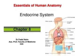
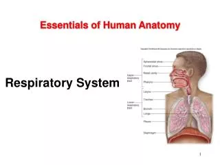
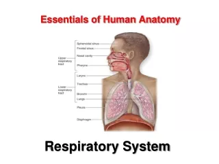
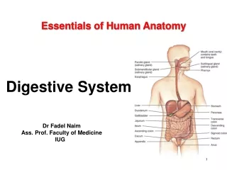
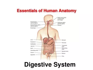
Essentials of Human Anatomy. Special Senses. Dr Fadel Naim Ass. Prof. Faculty of Medicine IUG. Special Senses. sensory receptors are within large, complex sensory organs in the head smell in olfactory organs taste in taste buds hearing and equilibrium in ears sight in eyes.

E N D
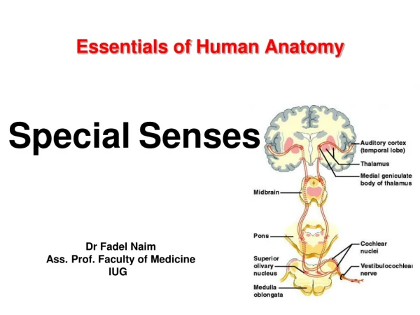
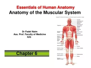
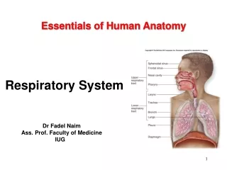
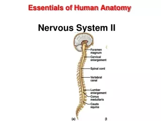
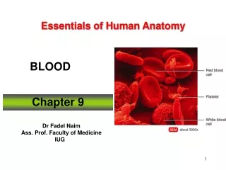
Essentials of Human Anatomy SpecialSenses Dr Fadel Naim Ass. Prof. Faculty of Medicine IUG
Special Senses • sensory receptors are within large, complex sensory organs in the head • smell in olfactory organs • taste in taste buds • hearing and equilibrium in ears • sight in eyes
Sense of Smell • Olfactory Receptors • chemoreceptors • respond to chemicals dissolved in liquids • Olfactory Organs • contain olfactory receptors and supporting epithelial cells • cover parts of nasal cavity, superior nasal conchae, and a portion of the nasal septum
Olfactory Nerve Pathways • Once olfactory receptors are stimulated, nerve impulses travel through • olfactory nerves olfactory bulbs olfactory tracts limbic system (for emotions) and olfactory cortex (for interpretation)
Olfactory Stimulation • olfactory organs located high in the nasal cavity above the usual pathway of inhaled air • olfactory receptors undergo sensory adaptation rapidly • sense of smell drops by 50% within a second after stimulation
Sense of Taste • Taste Buds • organs of taste • located on papillae of tongue, roof of mouth, linings of cheeks and walls of pharynx • Taste Receptors • chemoreceptors • taste cells – modified epithelial cells that function as receptors • taste hairs –microvilli that protrude from taste cells; sensitive parts of taste cells
Taste Sensations • Four Primary Taste Sensations • sweet – stimulated by carbohydrates • salty – stimulated by salts • sour – stimulated by acids • bitter – stimulated by many organic compounds Spicy foods activate pain receptors
Taste Nerve Pathways • Sensory impulses from taste receptors travel along • cranial nerves to • medulla oblongata to • thalamus to • gustatory cortex (for interpretation)
Hearing Ear – organ of hearing • Three Sections • External • Middle • Inner
External Ear • auricle • collects sounds waves • external auditory meatus • lined with ceruminous glands • carries sound to tympanic membrane • terminates with tympanic membrane • tympanic membrane • vibrates in response to sound waves
Middle Ear • tympanic cavity • air-filled space in temporal bone • auditory ossicles • vibrate in response to tympanic membrane • malleus, incus, and stapes • oval window • opening in wall of tympanic cavity • stapes vibrates against it to move fluids in inner ear
Auditory Tube • eustachian tube • connects middle ear to throat • helps maintain equal pressure on both sides of tympanic membrane • usually closed by valve-like flaps in throat
Inner Ear • complex system of labyrinths • osseous labyrinth • bony canal in temporal bone • filled with perilymph • membranous labyrinth • tube within osseous labyrinth • filled with endolymph
Inner Ear • Three Parts of Labyrinths • cochlea • functions in hearing • semicircular canals • functions in equilibrium • vestibule • functions in equilibrium
Cochlea • Scala vestibuli • upper compartment • leads from oval window to apex of spiral part of bony labyrinth • Scala tympani • lower compartment • extends from apex of the cochlea to round window • part of bony labyrinth
Cochlea • Cochlear duct • portion of membranous labyrinth in cochlea • Vestibular membrane • separates cochlear duct from scala vestibuli • Basilar membrane • separates cochlear duct from scala tympani
Organ of Corti • group of hearing receptor cells (hair cells) • on upper surface of basilar membrane • different frequencies of vibration move different parts of basilar membrane • particular sound frequencies cause hairs of receptor cells to bend • nerve impulse generated
Auditory Pathway Cochlear branch of CN VIII To inferior colliculus of opposite side of midbrain To thalamus To auditory cortex To cochlear nucleus of medulla
Vestibular Complex: • Semicircular canals with ampullae (mutually perpendicular) • Saccule and utricle (= fill up vestibule)
Two Receptor Organs:Maculae of Vestibule (or: macula of saccule plus macula of utricle)
Macula • responds to changes in head position • bending of hairs results in generation of nerve impulse
Crista Ampullaris • sensory organ of ampulla • hair cells and supporting cells • rapid turns of head or body stimulate hair cells
Equilibrium • Rotation of the head causes endolymph within the semicircular canal to push against the cupula covering the hair cells, resulting in bending of their stereocilia and the initiation of a nerve impulse.
Equilibrium • Dynamic Equilibrium • semicircular canals • sense rotation and movement of head and body • Static Equilibrium • vestibule • sense position of head when body is not moving
The Sense of Vision • Visual receptors (photoreceptors) in the eyes to detect light, color, and movement. • Accessory structures of the eye. • provide a superficial covering over its anterior exposed surface (conjunctiva) • prevent foreign objects from coming into contact with the eye (eyebrows, eyelashes, and eyelids) • keep the exposed surface moist, clean, and lubricated (lacrimal glands)
Lacrimal Apparatus • lacrimal gland • lateral to eye • secretes tears • canaliculi • collect tears • lacrimal sac • collects from canaliculi • nasolacrimal duct • collects from lacrimal sac • empties tears into nasal cavity
Structure of the Eye • hollow • spherical • wall has 3 layers • outer fibrous tunic • middle vascular tunic • inner nervous tunic
Cavities and Chambers of the Eye • The internal space of the eye is subdivided by the lens into two separate cavities. • anterior cavity • posterior cavity
Cavities and Chambers of the Eye • The anterior cavity is • the space anterior to the lens and posterior to the cornea • The iris of the eye subdivides the anterior cavity further into two chambers. • anterior chamber is between the iris and cornea • posterior chamber is between the lens and the iris
Vitreous Humor • Posterior cavity is posterior to the lens and anterior to the retina. • Transparent, gelatinous vitreous body which completely fills the space between the lens and the retina.
Optic Disc • Optic disc lacks photoreceptors. • Called the blind spot because no image forms there. • Just lateral to the optic disc is a rounded, yellowish region of the retina called the macula lutea containing a pit called the fovea centralis (the area of sharpest vision). • contains the highest proportion of cones and almost no rods
Visual Pathways • Each optic nerve conducts visual stimulus information. • At the optic chiasm, some axons from the optic nerve decussate. • The optic tract on each side then contains axons from both eyes. • Visual stimulus information is processed by the thalamus and then interpreted by visual association areas in the cerebrum.
Extrinsic Eye Muscles • Superior rectus • rotates eye up and medially • Lateral rectus • rotates eye laterally • Medial rectus • rotates eye medially • Inferior rectus • rotates eye down and medially • Superior oblique • rotates eye down and laterally • Inferior oblique • rotates eye up and laterally