Download
1 / 48
490 likes | 513 Vues
Essentials of Human Anatomy. Digestive System. General Structure of the Digestive System. Composed of two separate categories of organs: digestive organs accessory digestive organs. Digestive organs collectively make up the: gastrointestinal (GI) tract. Also called:

E N D
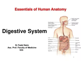
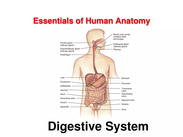
Essentials of Human Anatomy Digestive System
General Structure of the Digestive System • Composed of two separate categories of organs: • digestive organs • accessory digestive organs. • Digestive organs collectively make up the: • gastrointestinal (GI) tract. • Also called: • the digestive tract • alimentary canal.
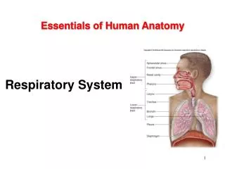
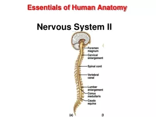
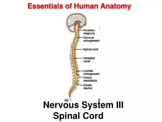
General Structure of the Digestive System • The GI tract organs: • oral cavity • pharynx • esophagus • stomach • small intestine • large intestine • continuous tube • about 30 feet (9–10 meters) • from mouth to anus. • Smooth muscle in the wall • responsible for motility • pushes materials from one end to the other.
General Structure of the Digestive System • Accessory digestive organs: • do not form the GI tube • are connected to the GI tract (some by ducts) • Assist the GI tract in the digestion of food. • Include: • Teeth • Tongue • Salivary glands • Liver • Gallbladder • Pancreas
Digestive System Functions • Ingestion • Digestion: break down of large particles of food • mechanical digestion • chemical digestion • Propulsion • peristalsis • segmentation • Secretion: • digestive enzymes • hormones • Absorption: • from external environment into internal environment • across mucosa • Elimination of wastes (defecation)
Oral Cavity (mouth) • Entrance to the GI tract. • Initial site of digestion: • mechanical digestion (via mastication) • chemical digestion (via enzymes in saliva). • Bounded anteriorly by the teeth and lips • Bounded posteriorly by the oropharynx. • Superior boundary is formed by the hard and soft palates. • Floor, or inferior surface, of the oral cavity • the tongue • the mylohyoid muscle covered with mucosa.
Oral Cavity (mouth) • Two regions of the oral cavity • Vestibule is the space between the cheeks or lips and the gums. • Oral cavity proper. • The lateral walls are formed by the cheeks. • Lips (labia). • Orbicularis oris muscle • Keratinized stratified squamous ET • Gingivae, or gums. • Dense regular CT • Nonkeratinized ET • Labial frenulum.
Palate • Hard palate • Anterior two-thirds of the palate • hard and bony • Soft palate • Posterior one-third • soft and muscular • primarily composed of skeletal muscle. • Extending inferiorly from the posterior part of the soft palate is the uvula. • When swallowing, the soft palate and the uvula elevate to close off the opening of the nasopharynx.
Tongue • An accessory digestive organ • Formed from: • skeletal muscle • covered with lightly keratinized stratified squamous epithelium. • Manipulates and mixes ingested materials during chewing • Forms the bolus. • a globular mass of partially digested material • Performs important functions in swallowing.
Tongue • Inferior surface of the tongue • attaches to the floor of the oral cavity • By the lingual frenulum. • Numerous small projections (papillae) cover the superior (dorsal) surface. • Posterior surface contains lingual tonsils. • Skeletal muscles move the tongue.
Salivary Glands • Collectively produce and secrete saliva. • a fluid that assists in the initial activities of digestion • Volume of saliva secreted daily ranges between 1.0 and 1.5 L. • Most is produced during mealtime • Smaller amounts are produced continuously to ensure that the oral cavity remains moist.
Salivary Glands • Components of saliva • Water: makes up 99% • Amylase: first step of chemical digestion • Lysozyme: antimicrobial • Functions • Moisten food • Food molecules into solution: taste • Form bolus: for swallowing • Cleanse oral cavity.
Salivary Glands • Three pairs of large, multicellular salivary glands: • parotid glands • submandibular glands • sublingual glands
The Parotid Glands • Largest salivary glands. • located anterior and inferior to the ear • partially overlying the masseter muscle. • Produce about 25–30% of saliva • conducted through the parotid duct to the oral cavity.
The Submandibular Glands • Inferior to the body of the mandible. • Produce most of the saliva (about 60–70%). • ducts opens through a papilla in the floor of the mouth • lateral to the the lingual frenulum.
The Sublingual Glands • Inferior to the tongue • internal to the oral cavity mucosa. • Each gland has multiple tiny sublingual ducts • open onto the inferior surface of the oral cavity • posterior to the submandibular duct papilla. • Contribute only about 3–5% of the total saliva.
Teeth • Two sets of teeth • 20 deciduous teeth, also called “milk teeth,” erupt between 6 months and 30 months after birth. • These teeth are eventually lost and replaced by 32 permanent teeth. • The more anteriorly placed permanent teeth tend to appear first, followed by the posteriorly placed teeth.
Teeth • The last teeth to erupt are the third molars, often called “wisdom teeth,” in the late teens or early 20’s.
General arrangement of abdominal GI organs • Peritoneum • Parietal peritoneum • Visceral peritoneum • Peritoneal cavity • Intraperitoneal organs • Retroperitoneal organs
Esophagus • Tubular passageway • Pharynx to stomach • Bolus • About 25 cm in adult • Esophageal hiatus: through diaphragm
Esophagus • Superior esophageal sphincter: • Skeletal muscle • Where pharynx and esophagus meet • Inferior esophageal sphincter • Also cardiac sphincter • Circular smooth muscle • Orifice between esophagus and stomach
Stomach • General • J-shaped • Functions • Digestion • Chemical • Mechanical • Results in chyme • Limited absorption
Stomach • Gross anatomy • Cardia • Cardiac orifice • Fundus • Body • Pylorus • Pyloric sphincter • Pyloric orifice • Greater curvature • Greater omentum • Lesser curvature • Lesser omemtum • Gastric folds
Stomach • Histology • Mucosa: simple columnar • Gastric pits • Gastric glands
Muscularis • 3 layers • Inner oblique • Middle circular • Outer longitudinal
Small Intestine • The duodenum • first segment of the small intestine. • approximately 25 centimeters (10 inches) long • originates at the pyloric sphincter • The jejunum • middle region of the small intestine. • approximately 2.5 meters (7.5 feet) • makes up approximately two-fifths of the small intestine’s total length. • primary region for chemical digestion and nutrient absorption • The ileum • is the last region of the small intestine. • about 3.6 meters (10.8 feet) in length • forms approximately three-fifths of the small intestine. • terminates at the ileocecal valve • sphincter that controls the entry of materials into the large intestine.
Large Intestine • approximate length of 1.5 meters (5 feet) • diameter of 6.5 centimeters (2.5 inches). • Absorbs most of the water and electrolytes from the remaining digested material. • Watery material that first enters the large intestine soon solidifies and becomes feces. • Stores fecal material until the body is ready to defecate. • Absorbs a very small percentage of nutrients still remaining in the digested material. • Composed of four segments: • the cecum, colon, rectum, anal canal
Accessory Digestive Organs • The liver • composed of four incompletely separated lobes • supported by two ligaments • Right lobe • Left lobe • Falciform ligament • Round ligament • Caudate lobe • Quadrate lobe
Functions of The Liver • Produce bile. • a greenish fluid that breaks down fats into small droplets to assist in their chemical digestion • Detoxify drugs, metabolites, and poisons. • Store excess nutrients and vitamins and release them when they are needed. • Synthesize blood plasmaproteins such as albumins, globulins, and proteins required for blood clotting. • Phagocytize debris in the blood. • Help break down and recycle components of aged erythrocytes and damaged or worn-out formed elements.
Accessory Digestive Organs • Gallbladder • concentrates bile produced by the liver and stores this concentrate until it is needed for digestion • cystic duct connects the gallbladder to the common bile duct • can hold approximately 40 to 60 milliliters of concentrated bile • Pancreas • mixed gland because it exhibits both endocrine and exocrine functions • Endocrine functions are performed by the pancreatic islets. • Exocrine activity results in the secretion of digestive enzymes, collectively called pancreatic juice, into the duodenum.
Accessory Digestive Organs • The biliary apparatus. • network of thin ducts that carry bile from the liver and gallbladder to the duodenum • the left and right lobes of the liver drain bile into the left andright hepatic ducts, respectively • the left and right hepatic ducts merge to form a single common hepatic duct • the cystic duct attaches to the common hepatic duct and carries bile to and from the gallbladder
Hepatitis • inflammation of the liver • most commonly caused by viral infection • can be caused by reactions to drug, alcoholism or autoimmunity • Signs and Symptoms • headache • low fever • fatigue • vomiting • rash • foamy urine • pale feces • jaundice • pain Hepatitis A – not washing hands or eating raw shellfish Hepatitis B – chronic; serum Hepatitis C – serum Hepatitis D – very severe; only produces symptoms if infected with B; serum HepatitisE, F, G – more rare