Download
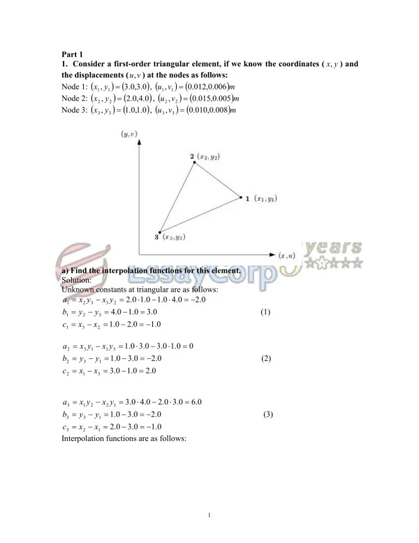
1 / 28
290 likes | 626 Vues
Medical image processing and finite element analysis. András Hajdu UNIVERSITY OF DEBRECEN HUNGARY. SSIP 2003 July 3-12 , 2003 Timişoara, Romania. MEDIP Platform independent software system for medical image processing.

E N D
Medical image processing and finite element analysis András Hajdu UNIVERSITY OF DEBRECEN HUNGARY SSIP 2003 July 3-12, 2003 Timişoara, Romania
MEDIPPlatformindependent software system for medical imageprocessing The aim of the project is to develop an informatical background to theoretical and applied studies in the field of multi-modal medical image processing,which results may lead to marketable products.
Developers Test partners • Department of Information Technology,University of Debrecen • PET Center, University of Debrecen • Mediso Medical Imaging System Ltd. • Department of Orthopedic Surgery,University of Debrecen • Faculty of Health Sciences, Chair of Radiotherapy,Semmelweis University • Faculty of Medicine Dept. of Radiology and Oncotherapy,Semmelweis University MEDIPPlatformindependent software system for medical imageprocessing
MEDIPPlatformindependent software system for medical imageprocessing 01.2003 01.2002 01.2004 • Survey, problem specification • Modelling, system plans • Implementation • Implementation,optimisation • Fine tuning,testing, presentation Pert diagram Dependence finished sessions current session Ses1 Ses2 Ses3 Ses4 Ses5 future sessions Feedback
MEDIP – Demostration programsPlatformindependent software system for medical imageprocessing Finite element modelling for virtual surgery Selection of volume of interest based on image fusion 4D visualization of gated heart and lung inspections
Demonstration program Finite element modelingfor virtual surgery Dept. of Information Tech., UD Dept. of Orthopaedy, UD
Demonstration program Finite element modelingfor virtual surgery Connecting to the base libraries • File I/O (DICOM) • Segmentation techniques • Contour tracking, ROI selection • Morphological operations • Complex GUI • ROI and VOI 2D/3D visualization • 3D geometric navigation • Printing
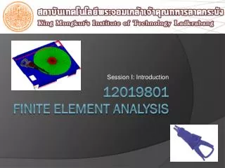
Demonstration program FEM surgery planning frame program Login (database opening) Launching (opening new/existing profile) DICOM file import Image manipulation (morphological filtering) Segmentation (automatic/manual) Creating geometric model
Demonstration program Surgery planning (virtual osteotomy) Adjusting parameters FEM contact 3D visualization, selecting VOI
Surgery planning (virtual osteotomy)Case study ANALYZIS OF A NEW FEMUR LEGTHENING SURGERY Zoltán Csernátony, Department of Orthopaedics, UD Szabolcs Molnár, Department of Orthopaedics, UD Sándor Manó, College Faculty of Engineering, UD AndrásHajdu, Institute of Informatics, UD ZoltánZörgő, Institute of Informatics, UD
Lowerextremityinequality Shorter femur Shorter tibia
Orthopaedic shoes Handling the problem (I.)
Surgical intervention(after Wagner and Ilizarov) distancing ossification Handling the problem (II.) cutting
A new lengthening method ! • Torsion and angulation could also be correnced
Past and present • In the past there existed no way of testing new interventions but to try it out in vivo • This days technology makes is possible to test and adjust new operative interventions before even one cut is made
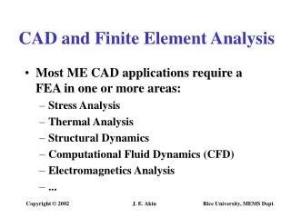
Validating new ideas • Laboratory tests • Finite element analysis
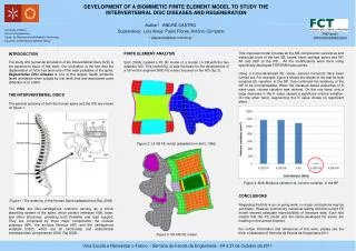
How can we use FEM/FEA? CT slices imageprocessing geometricalreconstruction
Importing images in CT firmware format (DICOM) Image enhancement (sharpening, filtering) Extracting ROIs Building up a basic model
Applying contour splines (Euclidean geometry) Reconstructing solid model (Coons patches) Building up a basic model
Based on the path of the cutting tool We need to determine: Cutting thickness Pitch Ending hole parameters Modeling intervention geometry (I.)
We subtract the object representing the „removed tissue” from the femur model Modeling interventiongeometry (II.)
The bone tissue should be modeled as a very complicated nonlinear anisotropic material We are using linearelasticizotropicandorthotropicmaterial models instead We mesh the model One end is fixed, on the other end a traction force is applied How big is the evolved stress? How much elongation can it support? Modeling intervention physics
Some results There exists an optimal combination of parameter values
Additional adjustments • The greatest stress values evolve near the ending holes. • The inward oriented conical bore appears to be the most suitable
We have built a schematic model It is a cylindrical pipe with inner and outer diameters equaling the femur’s average diameters The same analysis were performed Checking the results • The stress values measured on the pipe-model were 1.61 (D=0.27) times lower under same conditions • There was a 91% correlation between the two datasets
Future plans • More precise material modeling by using cylindrical layers, and other element types • In-vitro lab tests based on the results