Download
1 / 57
570 likes | 693 Vues
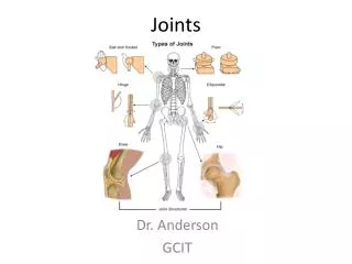
Lecture no 4. joints. Prepared by Dr.Salah Mohammad Fateh MBChB,DMRD,FIBMS(radiology). Types of arthritis. Degenerative arthritis (OA). Is the commonest form of arthritis. Changes occur secondary to wear & tear of the articular cartilage. Radiological features of OA.

E N D
Lecture no 4 joints Prepared by Dr.Salah Mohammad Fateh MBChB,DMRD,FIBMS(radiology)
Degenerative arthritis (OA) • Is the commonest form of arthritis. • Changes occur secondary to wear & tear of the articular cartilage.
OA • Joint space narrowed max. at wt bearing site • Erosion do no occur. • Subchondral sclerosis may be seen. • Sclerosis is a prominent feature. • No osteoporosis. • No peri articular soft tissue swelling • RA • Joint space narrowing uniform. • Erosin is characteristic feature. • Subchondral sclerosis is not a feature. • Sclerosis not a feature unless there is secondary OA. • Osteoporosis often present • Peri articular soft tissue swelling
OA RA
Most often due to pyogenic bacterial infection or TB. • Usually only one joint affected. • Synovial biopsy or exam. of the joint fluid is necessary for identification of infecting organism
Pyogenic infection • Usually due to staph. Aureus. • Rapid destruction of the articular cartilage followed by destruction of the subchondral bone & cause peri articual soft tissue swelling. • Earliest radiological finding is joint effusion, do US, you can do US guided aspiration of the joint fluid. • If Dx is still in doubt , then MRI advisable
There is decrease in cartilage width in the left hip, and cortical indistinctness in the left acetabulum with subarticular cyst formation.
TB arthritis • Hip& knee are the most commonly affected peripheral joints. • Spine involved in 50% of cases.
Radiological features • Localized osteoporosis. • Cartilage erosion usually occur late ,for that reason , at 1st joint space is preserved. • Marginal erosion. • At late stage there may be gross disorganization of the joint with calcified debris near the joint.
Also known as osteonecrosis, is where there is death of bone due to interruption of the blood supply. It occur most commonly in the intra-articular portions of bones & is associated with numerous underlying condition including. Steroid therapy. Collagen vascular diseases. Radiation therapy. Sickle cell disease. Exposure to the high pressure environment e.g. deep- see divers
X-ray finding Increased density of the subchondral bone with irregularity of the articular contour or even fragmentation A characteristic lucent line may be seen just beneath the articular cortex. The cartilage space may be preserved until secondary OA changes occur.
left hip joint; increased density centrally and flattening of the femoral head in the weight-bearing region, as well as the crescent sign or subchondral fracture.
MRI Is imaging modality of choice. It can show abnormality when the X-ray is normal & signal pattern allow specific Dx to be made.
The MR, shows that this patient has bilateral avascular necrosis of the hip joints, with a low-signal rim surrounding the necrotic segments
Perthe’s disease Is avascular necrosis of the femoral head in children. seen generally between ages 4 and 8, when the vascular supply to the femoral head is most at risk. Males are affected more than females. Bilateral in 10 percent of patients.
X-ray finding The first radiographic sign may be effusion. Later, increased density, fragmentation and flattening of the ossification center & lucent areas within it Metaphyseal irregularity & short wide femoral neck.
The left femoral capital epiphysis is dense, has lucent areas within it, and is flattened. This left hip is laterally subluxated,