Download
1 / 3
30 likes | 45 Vues
Usually, the right crus of the diaphragm attach onto the upper and the left crus onto the upper two lumbar vertebrae. In the present case, bilateral duplication of the diaphragmatic crura was noted. The right crus duplicated into right medial crus which had the usual attachment and right lateral crus that attached to the fifth lumbar vertebra. The left crus also duplicated into left medial crus which had the usual attachment and left lateral crus that attached to the fifth lumbar vertebra. A retrocrural space separated the two duplications.

E N D
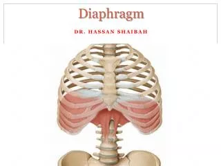
[AMJ 2019;12(2):53-55] Bilateral duplicated crura of the thoracoabdominal diaphragm Lydia S. Andrade1, Robert Kevin Fernandez1, and Antony Sylvan D’ souza2 1. Department of Anatomy, Kasturba Medical College, Manipal Academy of Higher Education, Manipal, Karnataka, 576104 India 2. DM Wayanad Institute of Medical Sciences, Naseera Nagar, Meppadi (P.O.), Wayanad, Kerala, 673577 India 3. What are the implications for research, policy, or practice? This case report explains the bilateral variation in the diaphragmatic crura so as to plan surgeries in the thoraco- abdominal region. CASE STUDY Please cite this paper as: Andrade LS, Fernandez RK, D’souza AS. Bilateral duplicated crura of the thoracoabdominal diaphragm. AMJ 2019;12(2):53–55. https://doi.org/10.21767/AMJ.2018.3529 Background The thoracoabdominal diaphragm is a musculo-aponeurotic partition between the thorax and the abdomen. It presents a sternal origin from the posterior surface of the Xiphisternum, a coastal origin from the lower six ribs, and a lumbar origin from the lumbar vertebrae. All these fibers insert into the central tendon. The lumbar origin is in the form of two crura and medial and lateral arcuate ligaments. The right crus is longer and stronger. It attaches to the anterior surface of the upper three lumbar vertebrae and the intervening intervertebral discs. The left crus is shorter and takes origin from the anterior surface of the bodies of the upper two lumbar vertebrae and the intervening intervertebral disc. Studies conducted in the past have shown that the lowest attachment of the right crus is to the fourth lumbar vertebra and that of the left crus is to the third lumbar vertebra.1,2 Studies have also highlighted the formation of the oesophageal hiatus from both the crura3 and its role on the gastroesophageal junction.4 Several reports have shown the unilateral duplication of right crus.5-7 This case report highlights the bilateral variation in the diaphragmatic crura with the embryological and clinical importance associated with the finding. Case details During the regular dissection for the undergraduate students, variation was noted in the attachments of both the crura of the diaphragm, in a 60-year-old female cadaver. The left crus duplicated to form a left medial crus (LMC) and a left lateral crus (LLC). A left retrocrural space (LRS) Corresponding Author: Lydia S. Andrade Department of Anatomy, Kasturba Medical College, MAHE, Manipal – 576104, Karnataka, India Email: lidibudy@gmail.com ABSTRACT Usually, the right crus of the diaphragm attach onto the upper and the left crus onto the upper two lumbar vertebrae. In the present case, bilateral duplication of the diaphragmatic crura was noted. The right crus duplicated into right medial crus which had the usual attachment and right lateral crus that attached to the fifth lumbar vertebra. The left crus also duplicated into left medial crus which had the usual attachment and left lateral crus that attached to the fifth lumbar vertebra. A retrocrural space separated the two duplications. A thorough knowledge of crural variations is necessary for the surgeons during surgical interventions. Key Words Crura, diaphragm, lumbar vertebrae Implications for Practice: 1. What is known about this subject? Unilateral variation of the diaphragmatic crus is more noted than bilateral variation. 2. What new information is offered in this case study? A rare bilateral variation in the origin of diaphragmatic crura. 53
[AMJ 2019;12(2):53-55] separated the two crura. The LMC attached to the anterior surface of the body of the second lumbar vertebra and merged with the anterior longitudinal ligament of the vertebrae. The LLC attached to the anterior surface of the body of the fifth lumbar vertebra. It sent additional slips of attachment to the anterior surface of the body of the fourth lumbar vertebra (Figure 1). Similarly, the right crus also presented a right medial crus (RMC) and a right lateral crus (RLC). A right retrocrural space (RRS) separated the two crura. The RMC attached to the anterior surface of the body of the third lumbar vertebra and merged with the anterior longitudinal ligament of the vertebrae. The RLC attached to the anterior surface of the body of the fifth lumbar vertebra (Figure 2). Splanchnic nerves passed through the left and right retrocrural spaces. The hemi-azygos vein (from the left renal vein) also passed through the LRS (Figure 3). Discussion Diaphragmatic variations may be due to alteration in its development. Formation of the diaphragm, in the intrauterine life, spans from four to twelve weeks. Diaphragm receives a contribution from the septum transversum, myoblasts arising from the 3rd, 4th and 5th cervical segments, pleuroperitoneal membranes and the dorsal mesentery of the esophagus. The crura are known to develop from the myoblasts that grow into the dorsal mesentery of the esophagus.8,9 One of the developmental anomalies that involve crura may be a partial duplication of the diaphragm. According to the standard anatomy description, the right crus attaches onto the bodies of the first three lumbar vertebrae and the left crus attaches onto the bodies of the first two lumbar vertebrae. However, studies conducted in the past have shown that the left crus could be longer and may attach onto the third1 and the right crus may attach onto the fourth lumbar vertebra.2 In the present case, both the crura had the usual attachment and an additional attachment to the fifth lumbar vertebra. The LLC sent additional slips to the fourth lumbar vertebra. Duplication of crura does not occur frequently. Unilateral duplication of the right crus has been shown earlier.5-7 The bilateral occurrence of duplicated crura is a very rare finding. Therefore, this case is unique, in having bilateral duplicated diaphragmatic crura. The diaphragm is the principal muscle of respiration. However, the crural fibers take part in the formation of oesophageal hiatus and thus act upon the gastroesophageal junction by forming the gastroesophageal reflux barrier10 and preventing the development of gastroesophageal reflux.4 The oesophageal hiatus is formed predominantly by both the right and left crura4 and predominantly by the right crus.11,12 In the present case of bilateral duplicated crura, the medial crura of both sides form the oesophageal hiatus. The crura of the diaphragm are related to numerous structures. Therefore, it is necessary to be known to all the possible anatomical variations of the diaphragm in order to treat the thoracoabdominal deformities effectively. Since the aortic opening is bounded by both the crura, the aorta may get compressed against the vertebral bodies. This would cause compression syndrome owing to lack of/decreased arterial supply.13 The crural attachments may also exert pressure on the lumbar arteries. Cases of transient and recurrent paraplegia due to compression of the second right lumbar artery by the diaphragmatic crus have been noted.14 Radiological and/or histological examination has confirmed the presence of tumors/cysts around the diaphragm. These could be benign or malignant. The most common benign tumors may be bronchogenic or mesothelial cysts. The most common primary malignant lesion is rhabdomyosarcoma.15 These may develop primarily in the diaphragm or secondarily, may spread from elsewhere via vascular channels, such as a metastatic lung or oesophageal malignancies, endometriosis, ovarian cancer, thymoma, mesothelioma, hepatic carcinoma, causing discomfort with breathing, hypoxemia, dysphagia or pain over the thoracic and abdominal regions.16 Thus surgical repair/resection turns out to be mandatory in these conditions. Therefore, the knowledge of variations of the crura would enlighten the surgeons to carefully perform surgery in this region. The retrocrural space may also involve in the spread of certain metastasis like a lipoma, vascular anomalies associated with the great vessels in this region, such as the aorta, inferior vena cava, and azygos venous system.17 In the present case, the retrocrural space case allowed the passage of splanchnic nerves, which usually pierces the diaphragmatic crura to enter the abdomen to form the coeliac plexus. This unusual course is of great importance to radiologists and anesthetists in performing the imaging- guided techniques for percutaneous blockade of the coeliac plexus.17 The splanchnic nerves, in the retrocrural space, would have been entrapped/compressed onto the posterior body wall during respiration, leading to abnormal oesophageal, gastric and intestinal functions causing an 54
[AMJ 2019;12(2):53-55] 14.Batt M, Rogopoulos A, Benchimol D, et al. Paraplegia with lumbar artery compression by the diaphragmatic crus. J Vasc Surg. 2008;48:1017–21. 15.Kim MP, Hofstetter WL. Tumors of the diaphragm. Thorac Surg Clin. 2009;19:521–9. 16.Weksler B, Ginsberg RJ. Tumors of the diaphragm. Chest Surg Clin N Am. 1998;8:441–7. 17.Restrepo CS, Eraso A, Ocazionez D, et al. The diaphragmatic crura and retrocrural space: normal imaging appearance, variants, and pathologic conditions. Radiographics. 2008;28:1289–305. ACKNOWLEDGEMENTS The authors are grateful to the teaching and non-teaching staff for their support PEER REVIEW Not commissioned. Externally peer reviewed. CONFLICTS OF INTEREST The authors declare that they have no competing interests. adverse effect on the gastro-oesophageal reflux. Therefore, the spinal manipulative therapy of the thoracolumbar spine could help to alleviate entrapments of the splanchnic nerves as they pass through the crura, promoting normal gastrointestinal function. Conclusion The finding in this case is of great clinical importance to the medical fraternity as it throws light on the presence of rare bilateral variation of the diaphragmatic crura. References 1.Ahmad I, Kaukab N, Ikram M, et al. Anatomical variations of diaphragmatic crura. J Rawalpindi Medical College. 2011;15:120–2. 2.Mazhar S, Mogotlane RA, Ahmad F. Crura of the diaphragm: variation in their anatomy. J Anatomy. 1988;188:499–500. 3.Skandalakis JE (ed). Skandalakis Surgical anatomy, the embryologic and anatomic basis of modern medicine. Athens: Paschalidis Medical Publications; 2005. 4.Mittal RK. The crural diaphragm, an external lower esophageal sphincter, Gastroenterology. 1993;105:740–7. 5.Sirasanagandla SR, Nayak SB, Bhat KMR, et al. Duplicated right crus of the diaphragm: a cadaveric case report. J Can Chiropr Assoc. 2014;58:39–44. 6.Rao TR, Rao S. Accessory right crus of the diaphragm-a case report. Case studies journal. 2016;5:1–3. 7.Vadgaonkar R, Murlimanju BV, Prabhu LV, et al. Accessory crus of diaphragm- a human morphological case report. J Morphol Sci. 2013;30:140–2. 8.Sadler TW. Langman’s Medical Embryology. 10th ed. Baltimore: Lippincott Williams & Wilkins, 2006. 9.Moore KL, Persaud TVN. Development of the diaphragm. In: Moore KL, Persaud TVN, eds. The developing human: clinically oriented embryology. 7th ed. Philadelphia: Saunders, 2003:192–7. 10.De Troyer A, Sampson M, Sigrist S, et al. Action of costal and crural parts of the diaphragm on the rib cage in dog. J Appl Physiol. 1982; 3:30–9. 11.Loukas M, Wartmann Ch, Tubbs RS, et al. Morphologic variation of the diaphragmatic crura: a correlation with pathologic processes of the esophageal hiatus?. Folia Morphol. 2008;67:273–9. 12.Botros KG, Bondok AA, Gabr OM, et al. Anatomical variations in the formation of the human esophageal hiatus. Anat Anz. 1990;171:193–9. 13.Petrella S, Prates JC. Celiac trunk compression syndrome. A review. Int J Morphol. 2006;4:429–36. a definitive study. 55