Understanding Monosynaptic Reflexes and Neuromuscular Junction Dynamics
This resource explores the fundamentals of monosynaptic reflexes and the intricate processes at the neuromuscular junction (NMJ). It covers classical elements of synaptic transmission, focusing on transmitter release, synaptic currents, potentials, and integration. Key topics include the mechanics of exocytosis, presynaptic calcium channels, and the nature of neurotransmitter receptors (ionotropic and metabotropic). The document also emphasizes the importance of temporal and spatial summation in synaptic efficacy and the role of synaptic location affecting transmission dynamics.
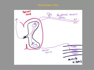

Understanding Monosynaptic Reflexes and Neuromuscular Junction Dynamics
E N D
Presentation Transcript
Amy MacDermott, Department of Physiology and Cellular Biophysics and the Center for Neurobiology and Behavior phone 305-3889 email abm1@columbia.edu Physiology G6001 Nerve and Synapse Classical elements of synaptic transmission: Neuromuscular junction Transmitter release Synaptic currents Synaptic potentials Nerve-nerve synapses Synaptic integration Summation Facilitation
Physiology G6001 Nerve and Synapse Classical elements of synaptic transmission: Neuromuscular junction Transmitter release Synaptic currents Synaptic potentials Nerve-nerve synapses Synaptic integration Summation Facilitation
Protein machinery for vesicle release Rizo and Sudhof Nature Reviews Neuroscience3; 641-653 (2002);
Recording from the neuromuscular junction (NMJ).Spontaneous release of a vesicle of Ach causes a miniature endplate potential or MEPPEvoked release following stimulation of the motor neuron causes an endplate potential or EPP
Presynaptic calcium channels at the neuromuscular junction (NMJ)
Pre and postsynaptic changes in membrane potential during transmitter release
Recording from the neuromuscular junction (NMJ).Spontaneous release of a vesicle of Ach causes a miniature endplate potential or MEPPEvoked release following stimulation of the motor neuron causes an endplate potential or EPP
The quantal nature of transmitter release.Decrease the amplitude of evoked release by recording in low Ca2+ bath.
Physiology G6001 Nerve and Synapse Classical elements of synaptic transmission: Neuromuscular junction Transmitter release Synaptic currents Synaptic potentials Nerve-nerve synapses Synaptic integration Summation Facilitation
NMJ – an inward current drives the change in membrane potential
Membrane potential and driving force– brief reviewVm= (RT/F) ln [K]o/[K]i Vm= (RT/F) ln Na]o/[Na]i
ACh binds to the nicotinic ACh receptor, causing it to gate open. The channel is permeable to both Na+ and K+.The end-plate potential causes voltage gated Na+ channels to open and an action potential to fire.
An EPP in normal muscle is super-threshold for firing action potential
Physiology G6001 Nerve and Synapse Classical elements of synaptic transmission: Neuromuscular junction Transmitter release Synaptic currents Synaptic potentials Nerve-nerve synapses Synaptic integration Summation Facilitation
a b - - - - - - - - - - - - - - - - - - - - - - - - + + + + + + g + + + + + + + + + + + + + + + + + Classes of neurotransmitter receptors - OUT + + + + - - - - - - - - - - - - IN IONOTROPIC METABOTROPIC
GLU GLU, KA GLU, NMDA b g Excitatory synaptic transmission is mediate by glutamate receptors (Ca2+?) Na+, Ca2+ Na+, GLU, KA OUT a IN * K+ K+ metabotropic Glu receptors NMDA receptors (NMDARs) AMPA receptors Kainate receptors
Current-voltage relationship for synaptic currents mediated by AMPA and NMDA receptors
Inhibitory synaptic transmission is mediated by GABA and glycine receptors
Physiology G6001 Nerve and Synapse Classical elements of synaptic transmission: Neuromuscular junction Transmitter release Synaptic currents Synaptic potentials Nerve-nerve synapses Synaptic integration Summation Facilitation
Temporal summation depends on the passive membrane properties of the neuron or muscle Tau or t = C x R
Summation is postsynaptic while facilitation is usually presynaptic
The synaptic potential is not actively propagated. The rate of decay with distance is exponential: DV(x) = DV0 e-x/l and l ~ (rm/ra)
Different synaptic configurations including axo-somatic, axodendritic and axo-axonic
Implications of synapse location Length constant l ~ Rm/Ra
Temporal and spatial summation: importance of time constant t and length constant l
Three forms of modulation of synaptic transmission mediated by metabotropic receptors
Berne and Levy – chapter 4orKandel, Schwartz, and Jessell – chapters 11 and 12or Kandel, Schwartz, and Jessell – chapters 10-15
Recording from the neuromuscular junction (NMJ).Spontaneous release of a vesicle of Ach causes a miniature endplate potential or MEPPEvoked release following stimulation of the motor neuron causes an endplate potential or EPP
The reversal potential is determined by the concentrations of ions flowing through the synaptic channel.